
- Science Notes Posts
- Contact Science Notes
- Todd Helmenstine Biography
- Anne Helmenstine Biography
- Free Printable Periodic Tables (PDF and PNG)
- Periodic Table Wallpapers
- Interactive Periodic Table
- Periodic Table Posters
- Science Experiments for Kids
- How to Grow Crystals
- Chemistry Projects
- Fire and Flames Projects
- Holiday Science
- Chemistry Problems With Answers
- Physics Problems
- Unit Conversion Example Problems
- Chemistry Worksheets
- Biology Worksheets
- Periodic Table Worksheets
- Physical Science Worksheets
- Science Lab Worksheets
- My Amazon Books

Parts of the Brain and Their Functions

The human brain is the epicenter of our nervous system and plays a pivotal role in virtually every aspect of our lives. It’s a complex, highly organized organ responsible for thoughts, feelings, actions, and interactions with the world around us. Here is a look at the intricate anatomy of the brain, its functions, and the consequences of damage to different areas.
Introduction to the Brain and Its Functions
The brain is an organ of soft nervous tissue that is protected within the skull of vertebrates. It functions as the coordinating center of sensation and intellectual and nervous activity. The brain consists of billions of neurons (nerve cells) that communicate through intricate networks. The primary functions of the brain include processing sensory information, regulating bodily functions, forming thoughts and emotions, and storing memories.
Main Parts of the Brain – Anatomy
The three main parts of the brain are the cerebrum, cerebellum, and brainstem.
1. Cerebrum
- Location: The cerebellum occupies the upper part of the cranial cavity and is the largest part of the human brain.
- Functions: It’s responsible for higher brain functions, including thought, action, emotion, and interpretation of sensory data.
- Effects of Damage: Depending on the area affected, damage leads to memory loss, impaired cognitive skills, changes in personality, and loss of motor control.
2. Cerebellum
- Location: The cerebellum is at the back of the brain, below the cerebrum.
- Functions: It coordinates voluntary movements such as posture, balance, coordination, and speech.
- Effects of Damage: Damage causes problems with balance, movement, and muscle coordination (ataxia).
3. Brainstem
- Location: The brainstem is lower extension of the brain, connecting to the spinal cord. It includes the midbrain, pons, and medulla oblongata.
- Functions: This part of the brain controls many basic life-sustaining functions, including heart rate, breathing, sleeping, and eating.
- Effects of Damage: Damage results in life-threatening conditions like breathing difficulties, heart problems, and loss of consciousness.
Lobes of the Brain
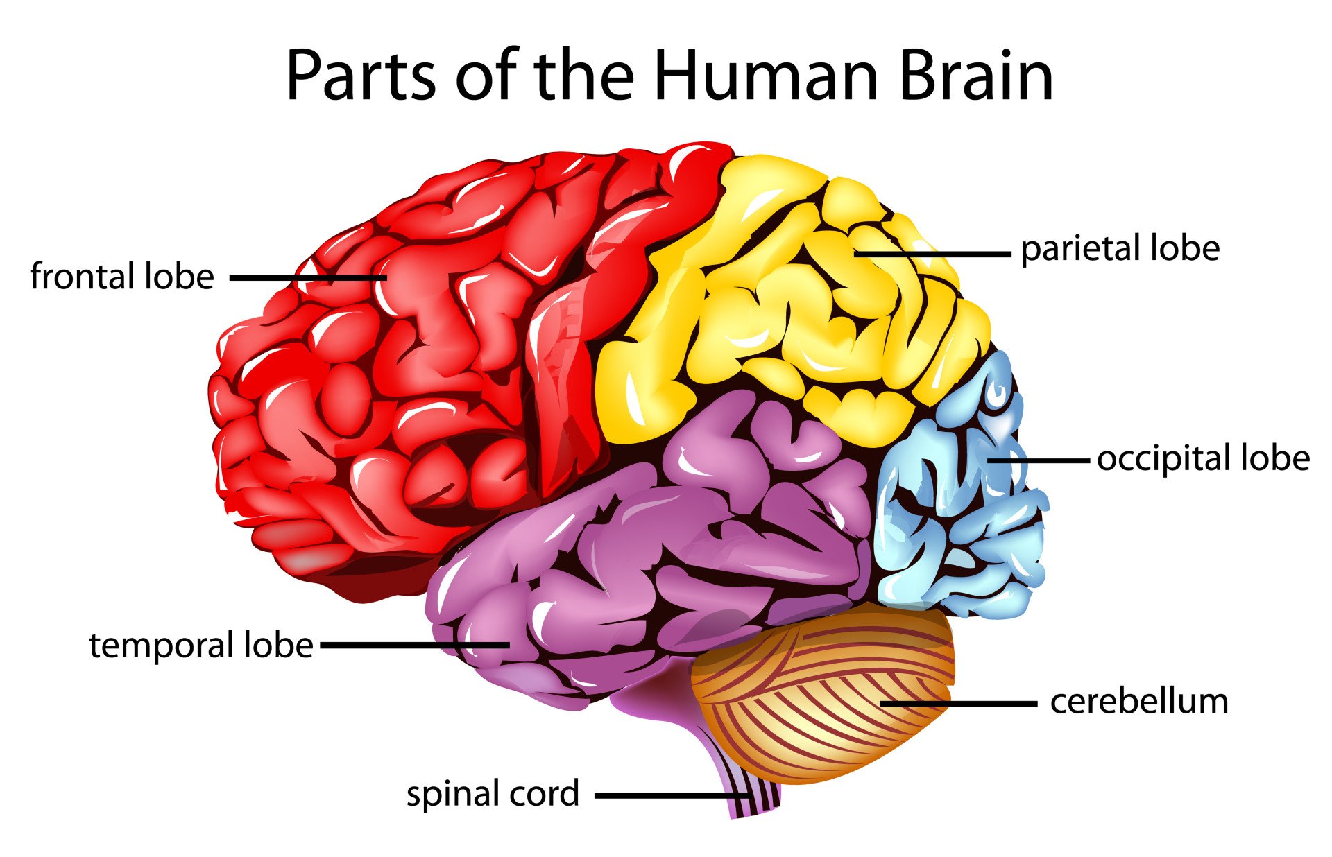
The four lobes of the brain are regions of the cerebrum:
- Location: This is the anterior or front part of the brain.
- Functions: Decision making, problem solving, control of purposeful behaviors, consciousness, and emotions.
- Location: Sits behind the frontal lobe.
- Functions: Processes sensory information it receives from the outside world, mainly relating to spatial sense and navigation (proprioception).
- Location: Below the lateral fissure, on both cerebral hemispheres.
- Functions: Mainly revolves around auditory perception and is also important for the processing of both speech and vision (reading).
- Location: At the back of the brain.
- Functions: Main center for visual processing.
Left vs. Right Brain Hemispheres
The cerebrum has two halves, called hemispheres. Each half controls functions on the opposite side of the body. So, the left hemisphere controls muscles on the right side of the body, and vice versa. But, the functions of the two hemispheres are not entirely identical:
- Left Hemisphere: It’s dominant in language and speech and plays roles in logical thinking, analysis, and accuracy. .
- Right Hemisphere: This hemisphere is more visual and intuitive and functions in creative and imaginative tasks.
The corpus callosum is a band of nerves that connect the two hemispheres and allow communication between them.
Detailed List of Parts of the Brain
While knowing the three key parts of the brain is a good start, the anatomy is quite a bit more complex. In addition to nervous tissues, the brain also contains key glands:
- Cerebrum: The cerebrum is the largest part of the brain. Divided into lobes, it coordinates thought, movement, memory, senses, speech, and temperature.
- Corpus Callosum : A broad band of nerve fibers joining the two hemispheres of the brain, facilitating interhemispheric communication.
- Cerebellum : Coordinates movement and balance and aids in eye movement.
- Pons : Controls voluntary actions, including swallowing, bladder function, facial expression, posture, and sleep.
- Medulla oblongata : Regulates involuntary actions, including breathing, heart rhythm, as well as oxygen and carbon dioxide levels.
- Limbic System : Includes the amygdala, hippocampus, and parts of the thalamus and hypothalamus.
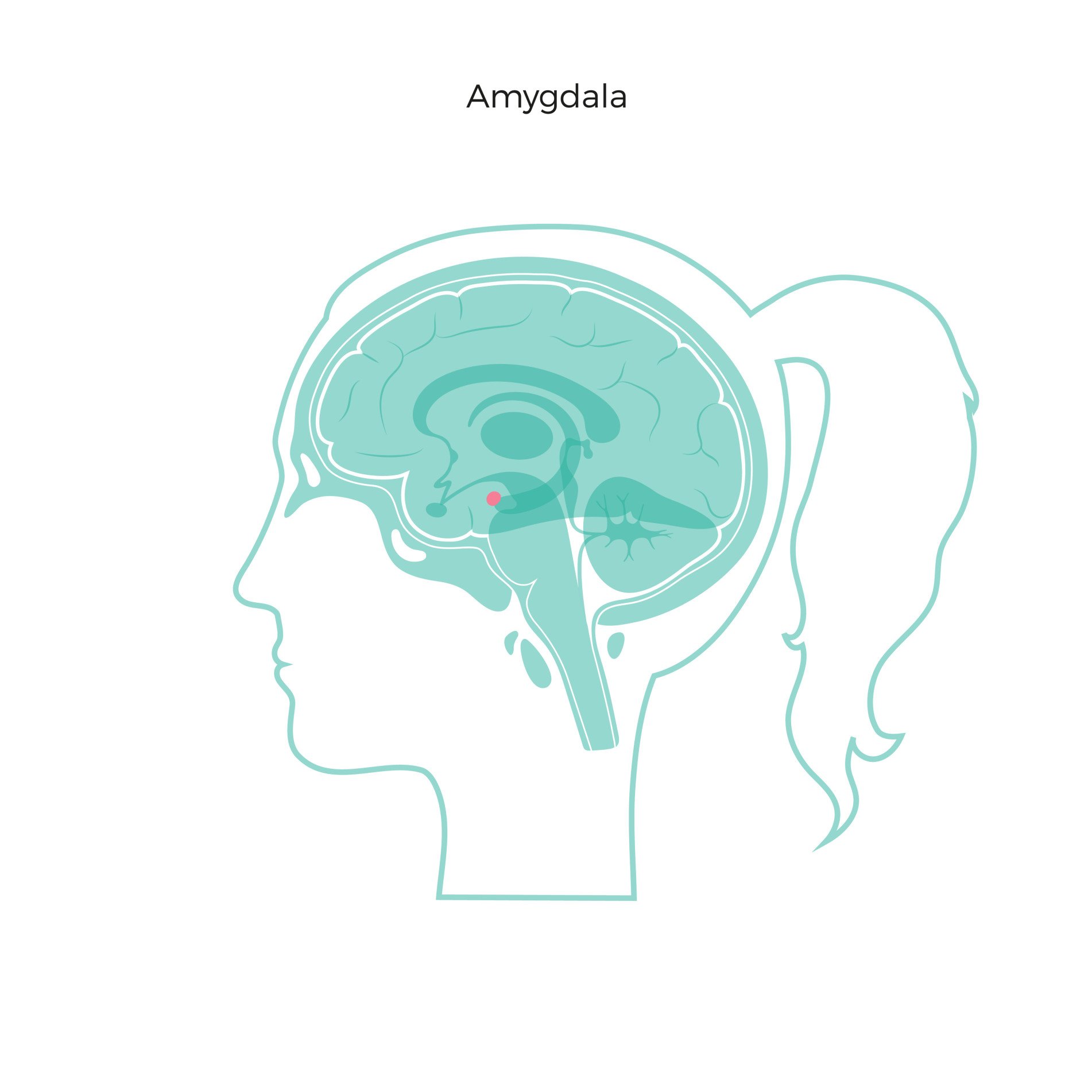
- Amygdala: Plays a key role in emotional responses, hormonal secretions, and memory formation.
- Hippocampus: Plays a vital role in memory formation and spatial navigation.
- Thalamus : Acts as the brain’s relay station, channeling sensory and motor signals to the cerebral cortex, and regulating consciousness, sleep, and alertness.
- Basal Ganglia : A group of structures involved in processing information related to movement, emotions, and reward. Key structures include the striatum, globus pallidus, substantia nigra, and subthalamic nucleus.
- Ventral Tegmental Area (VTA) : Plays a role in the reward circuit of the brain, releasing dopamine in response to stimuli indicating a reward.
- Optic tectum : Also known as the superior colliculus, it directs eye movements.
- Substantia Nigra : Involved in motor control and contains a large concentration of dopamine-producing neurons.
- Cingulate Gyrus : Plays a role in processing emotions and behavior regulation. It also helps regulate autonomic motor function.
- Olfactory Bulb : Involved in the sense of smell and the integration of olfactory information.
- Mammillary Bodies : Plays a role in recollective memory.
- Function: Regulates emotions, memory, and arousal.
Glands in the Brain
The hypothalamus, pineal gland, and pituitary gland are the three endocrine glands within the brain:
- Hypothalamus : The hypothalamus links the nervous and endocrine systems. It contains many small nuclei. In addition to participating in eating and drinking, sleeping and waking, it regulates the endocrine system via the pituitary gland. It maintains the body’s homeostasis, regulating hunger, thirst, response to pain, levels of pleasure, sexual satisfaction, anger, and aggressive behavior.
- Pituitary Gland : Known as the “master gland,” it controls various other hormone glands in the body, such as the thyroid and adrenals, as well as regulating growth, metabolism, and reproductive processes.
- Pineal Gland : The pineal gland produces and regulates some hormones, including melatonin, which is crucial in regulating sleep patterns and circadian rhythms.
Gray Matter vs. White Matter
The brain and spinal cord consist of gray matter (substantia grisea) and white matter (substantia alba).
- White Matter: Consists mainly of axons and myelin sheaths that send signals between different brain regions and between the brain and spinal cord.
- Gray Matter: Consists of neuronal cell bodies, dendrites, and axon terminals. Gray matter processes information and directs stimuli for muscle control, sensory perception, decision making, and self-control.
Frequently Asked Questions (FAQs) About the Human Brain
- The human brain contains approximately 86 billion neurons. Additionally, it has a similar or slightly higher number of non-neuronal cells (glial cells), making the total number of cells in the brain close to 170 billion.
- There are about 86 billion neurons in the human brain. These neurons are connected by trillions of synapses, forming a complex networks.
- The average adult human brain weighs about 1.3 to 1.4 kilograms (about 3 pounds). This weight represents about 2% of the total body weight.
- The brain is about 73% water.
- The myth that humans only use 10% of their brain is false. Virtually every part gets use, and most of the brain is active all the time, even during sleep.
- The average size of the adult human brain is about 15 centimeters (6 inches) in length, 14 centimeters (5.5 inches) in width, and 9 centimeters (3.5 inches) in height.
- Brain signal speeds vary depending on the type of neuron and the nature of the signal. They travel anywhere from 1 meter per second to over 100 meters per second in the fastest neurons.
- With age, the brain’s volume and/or weight decrease, synaptic connections reduce, and there can be a decline in cognitive functions. However, the brain to continues adapting and forming new connections throughout life.
- The brain has a limited ability to repair itself. Neuroplasticity aids recovery by allowing other parts of the brain to take over functions of the damaged areas.
- The brain consumes about 20% of the body’s total energy , despite only making up about 2% of the body’s total weight . It requires a constant supply of glucose and oxygen.
- Sleep is crucial for brain health. It aids in memory consolidation, learning, brain detoxification, and the regulation of mood and cognitive functions.
- Douglas Fields, R. (2008). “White Matter Matters”. Scientific American . 298 (3): 54–61. doi: 10.1038/scientificamerican0308-54
- Kandel, Eric R.; Schwartz, James Harris; Jessell, Thomas M. (2000). Principles of Neural Science (4th ed.). New York: McGraw-Hill. ISBN 978-0-8385-7701-1.
- Kolb, B.; Whishaw, I.Q. (2003). Fundamentals of Human Neuropsychology (5th ed.). New York: Worth Publishing. ISBN 978-0-7167-5300-1.
- Rajmohan, V.; Mohandas, E. (2007). “The limbic system”. Indian Journal of Psychiatry . 49 (2): 132–139. doi: 10.4103/0019-5545.33264
- Shepherd, G.M. (1994). Neurobiology . Oxford University Press. ISBN 978-0-19-508843-4.
Related Posts
Masks Strongly Recommended but Not Required in Maryland, Starting Immediately
Due to the downward trend in respiratory viruses in Maryland, masking is no longer required but remains strongly recommended in Johns Hopkins Medicine clinical locations in Maryland. Read more .
- Vaccines
- Masking Guidelines
- Visitor Guidelines

Brain Anatomy and How the Brain Works
What is the brain.
The brain is a complex organ that controls thought, memory, emotion, touch, motor skills, vision, breathing, temperature, hunger and every process that regulates our body. Together, the brain and spinal cord that extends from it make up the central nervous system, or CNS.
What is the brain made of?
Weighing about 3 pounds in the average adult, the brain is about 60% fat. The remaining 40% is a combination of water, protein, carbohydrates and salts. The brain itself is a not a muscle. It contains blood vessels and nerves, including neurons and glial cells.
What is the gray matter and white matter?
Gray and white matter are two different regions of the central nervous system. In the brain, gray matter refers to the darker, outer portion, while white matter describes the lighter, inner section underneath. In the spinal cord, this order is reversed: The white matter is on the outside, and the gray matter sits within.

Gray matter is primarily composed of neuron somas (the round central cell bodies), and white matter is mostly made of axons (the long stems that connects neurons together) wrapped in myelin (a protective coating). The different composition of neuron parts is why the two appear as separate shades on certain scans.
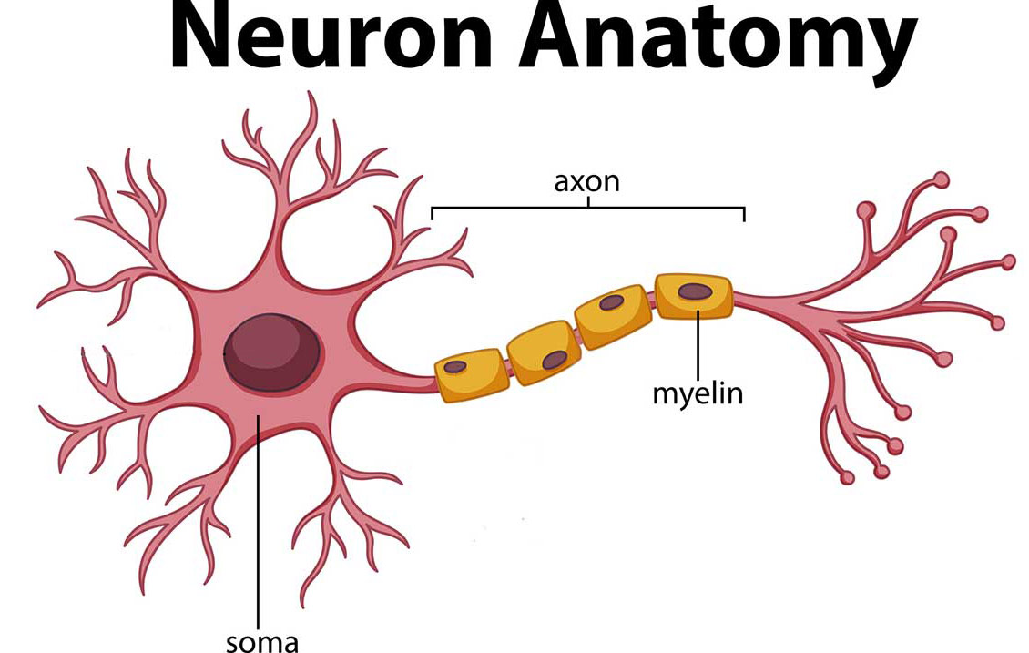
Each region serves a different role. Gray matter is primarily responsible for processing and interpreting information, while white matter transmits that information to other parts of the nervous system.
How does the brain work?
The brain sends and receives chemical and electrical signals throughout the body. Different signals control different processes, and your brain interprets each. Some make you feel tired, for example, while others make you feel pain.
Some messages are kept within the brain, while others are relayed through the spine and across the body’s vast network of nerves to distant extremities. To do this, the central nervous system relies on billions of neurons (nerve cells).
Main Parts of the Brain and Their Functions
At a high level, the brain can be divided into the cerebrum, brainstem and cerebellum.
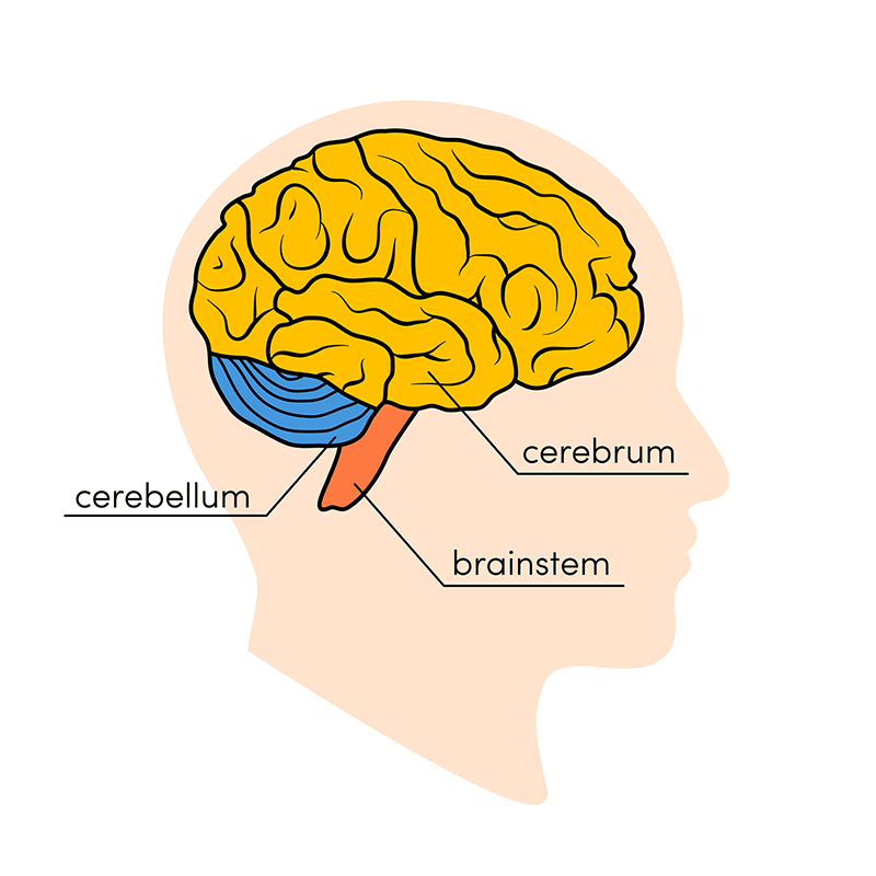
The cerebrum (front of brain) comprises gray matter (the cerebral cortex) and white matter at its center. The largest part of the brain, the cerebrum initiates and coordinates movement and regulates temperature. Other areas of the cerebrum enable speech, judgment, thinking and reasoning, problem-solving, emotions and learning. Other functions relate to vision, hearing, touch and other senses.
Cerebral Cortex
Cortex is Latin for “bark,” and describes the outer gray matter covering of the cerebrum. The cortex has a large surface area due to its folds, and comprises about half of the brain’s weight.
The cerebral cortex is divided into two halves, or hemispheres. It is covered with ridges (gyri) and folds (sulci). The two halves join at a large, deep sulcus (the interhemispheric fissure, AKA the medial longitudinal fissure) that runs from the front of the head to the back. The right hemisphere controls the left side of the body, and the left half controls the right side of the body. The two halves communicate with one another through a large, C-shaped structure of white matter and nerve pathways called the corpus callosum. The corpus callosum is in the center of the cerebrum.
The brainstem (middle of brain) connects the cerebrum with the spinal cord. The brainstem includes the midbrain, the pons and the medulla.
- Midbrain. The midbrain (or mesencephalon) is a very complex structure with a range of different neuron clusters (nuclei and colliculi), neural pathways and other structures. These features facilitate various functions, from hearing and movement to calculating responses and environmental changes. The midbrain also contains the substantia nigra, an area affected by Parkinson’s disease that is rich in dopamine neurons and part of the basal ganglia, which enables movement and coordination.
- Pons. The pons is the origin for four of the 12 cranial nerves, which enable a range of activities such as tear production, chewing, blinking, focusing vision, balance, hearing and facial expression. Named for the Latin word for “bridge,” the pons is the connection between the midbrain and the medulla.
- Medulla. At the bottom of the brainstem, the medulla is where the brain meets the spinal cord. The medulla is essential to survival. Functions of the medulla regulate many bodily activities, including heart rhythm, breathing, blood flow, and oxygen and carbon dioxide levels. The medulla produces reflexive activities such as sneezing, vomiting, coughing and swallowing.
The spinal cord extends from the bottom of the medulla and through a large opening in the bottom of the skull. Supported by the vertebrae, the spinal cord carries messages to and from the brain and the rest of the body.
The cerebellum (“little brain”) is a fist-sized portion of the brain located at the back of the head, below the temporal and occipital lobes and above the brainstem. Like the cerebral cortex, it has two hemispheres. The outer portion contains neurons, and the inner area communicates with the cerebral cortex. Its function is to coordinate voluntary muscle movements and to maintain posture, balance and equilibrium. New studies are exploring the cerebellum’s roles in thought, emotions and social behavior, as well as its possible involvement in addiction, autism and schizophrenia.
Brain Coverings: Meninges
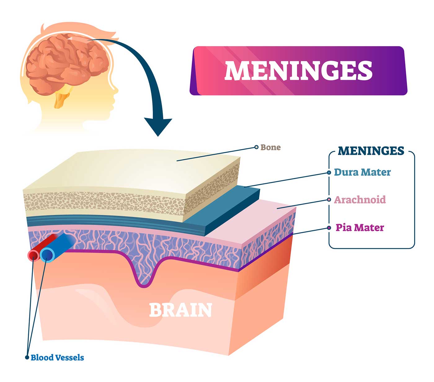
Three layers of protective covering called meninges surround the brain and the spinal cord.
- The outermost layer, the dura mater , is thick and tough. It includes two layers: The periosteal layer of the dura mater lines the inner dome of the skull (cranium) and the meningeal layer is below that. Spaces between the layers allow for the passage of veins and arteries that supply blood flow to the brain.
- The arachnoid mater is a thin, weblike layer of connective tissue that does not contain nerves or blood vessels. Below the arachnoid mater is the cerebrospinal fluid, or CSF. This fluid cushions the entire central nervous system (brain and spinal cord) and continually circulates around these structures to remove impurities.
- The pia mater is a thin membrane that hugs the surface of the brain and follows its contours. The pia mater is rich with veins and arteries.
Lobes of the Brain and What They Control
Each brain hemisphere (parts of the cerebrum) has four sections, called lobes: frontal, parietal, temporal and occipital. Each lobe controls specific functions.
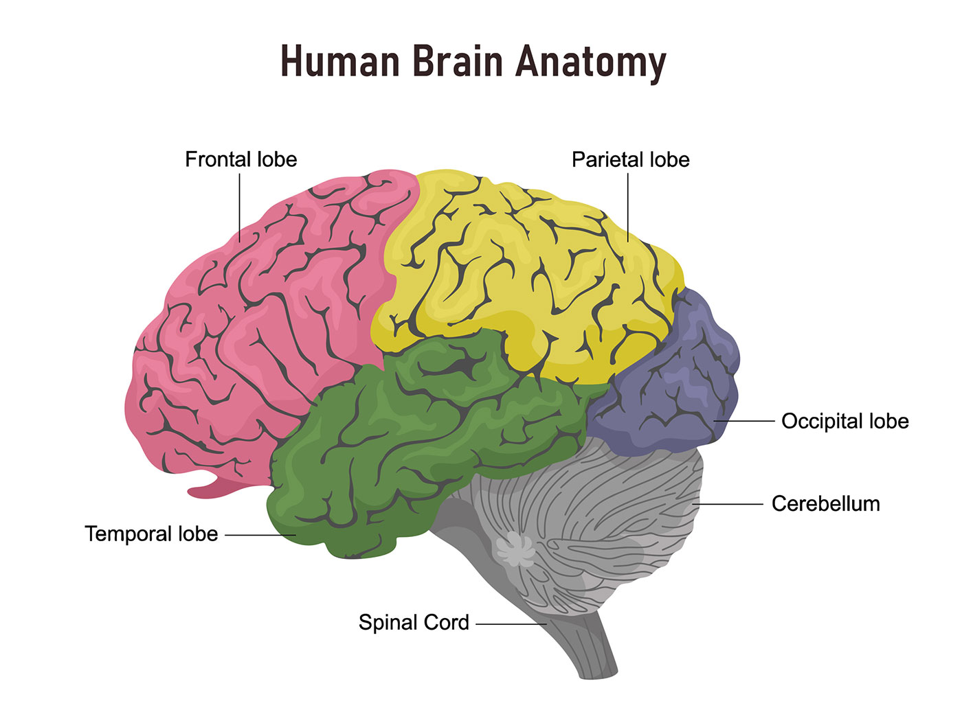
- Frontal lobe. The largest lobe of the brain, located in the front of the head, the frontal lobe is involved in personality characteristics, decision-making and movement. Recognition of smell usually involves parts of the frontal lobe. The frontal lobe contains Broca’s area, which is associated with speech ability.
- Parietal lobe. The middle part of the brain, the parietal lobe helps a person identify objects and understand spatial relationships (where one’s body is compared with objects around the person). The parietal lobe is also involved in interpreting pain and touch in the body. The parietal lobe houses Wernicke’s area, which helps the brain understand spoken language.
- Occipital lobe. The occipital lobe is the back part of the brain that is involved with vision.
- Temporal lobe. The sides of the brain, temporal lobes are involved in short-term memory, speech, musical rhythm and some degree of smell recognition.
Deeper Structures Within the Brain
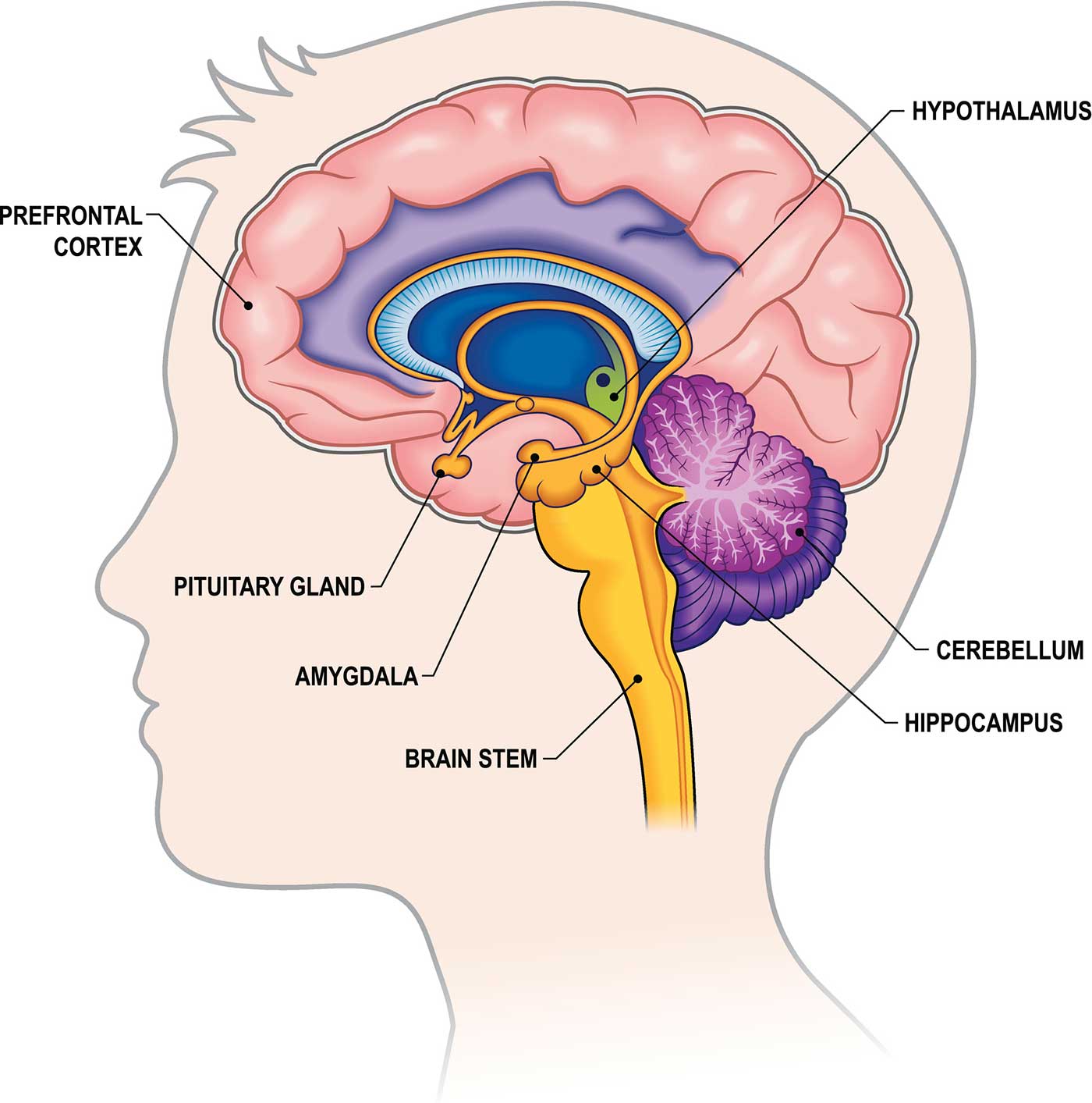
Pituitary gland.
Sometimes called the “master gland,” the pituitary gland is a pea-sized structure found deep in the brain behind the bridge of the nose. The pituitary gland governs the function of other glands in the body, regulating the flow of hormones from the thyroid, adrenals, ovaries and testicles. It receives chemical signals from the hypothalamus through its stalk and blood supply.
Hypothalamus
The hypothalamus is located above the pituitary gland and sends it chemical messages that control its function. It regulates body temperature, synchronizes sleep patterns, controls hunger and thirst and also plays a role in some aspects of memory and emotion.
Small, almond-shaped structures, an amygdala is located under each half (hemisphere) of the brain. Included in the limbic system, the amygdalae regulate emotion and memory and are associated with the brain’s reward system, stress, and the “fight or flight” response when someone perceives a threat.
Hippocampus
A curved seahorse-shaped organ on the underside of each temporal lobe, the hippocampus is part of a larger structure called the hippocampal formation. It supports memory, learning, navigation and perception of space. It receives information from the cerebral cortex and may play a role in Alzheimer’s disease.
Pineal Gland
The pineal gland is located deep in the brain and attached by a stalk to the top of the third ventricle. The pineal gland responds to light and dark and secretes melatonin, which regulates circadian rhythms and the sleep-wake cycle.
Ventricles and Cerebrospinal Fluid
Deep in the brain are four open areas with passageways between them. They also open into the central spinal canal and the area beneath arachnoid layer of the meninges.
The ventricles manufacture cerebrospinal fluid , or CSF, a watery fluid that circulates in and around the ventricles and the spinal cord, and between the meninges. CSF surrounds and cushions the spinal cord and brain, washes out waste and impurities, and delivers nutrients.
Blood Supply to the Brain
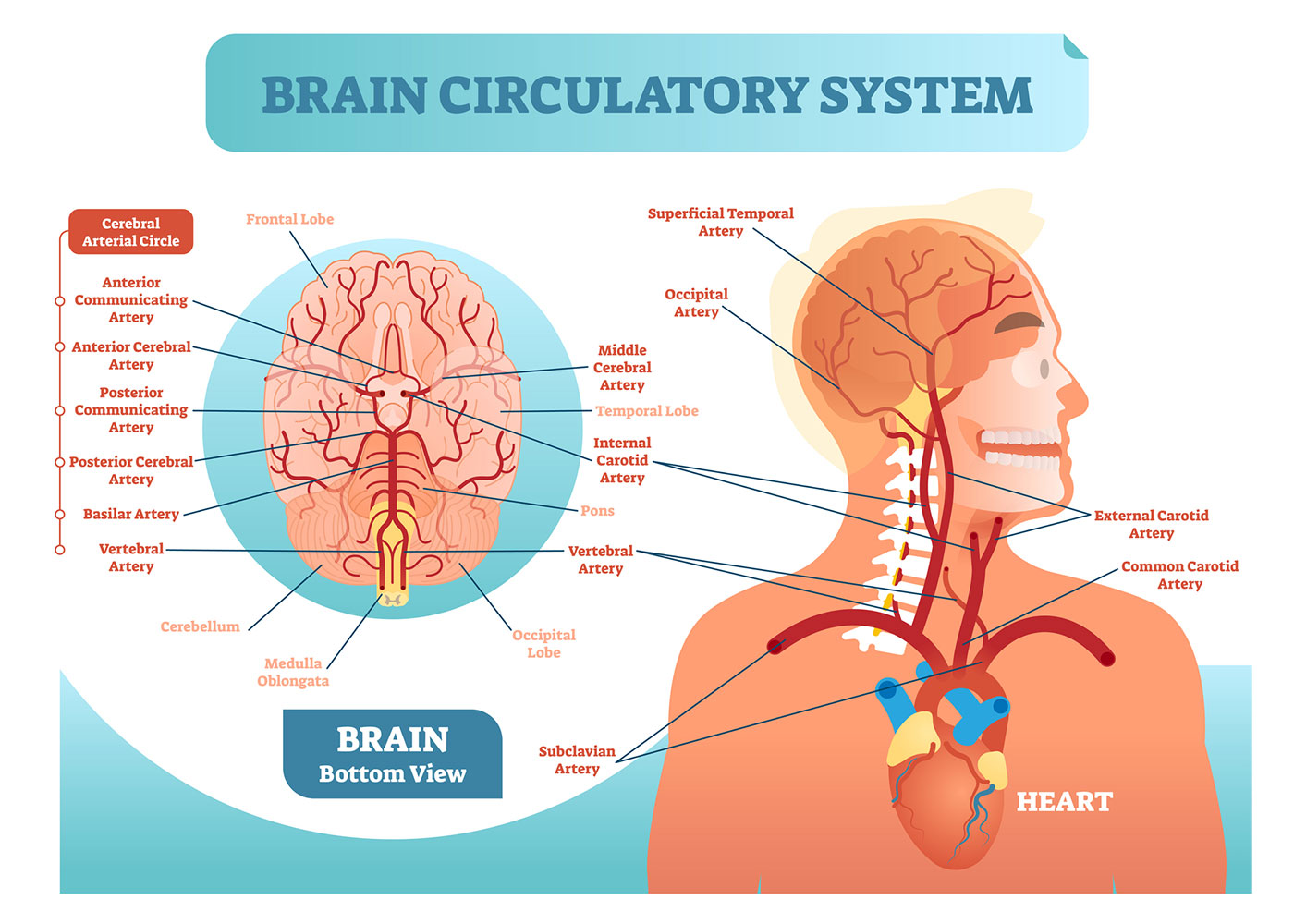
Two sets of blood vessels supply blood and oxygen to the brain: the vertebral arteries and the carotid arteries.
The external carotid arteries extend up the sides of your neck, and are where you can feel your pulse when you touch the area with your fingertips. The internal carotid arteries branch into the skull and circulate blood to the front part of the brain.
The vertebral arteries follow the spinal column into the skull, where they join together at the brainstem and form the basilar artery , which supplies blood to the rear portions of the brain.
The circle of Willis , a loop of blood vessels near the bottom of the brain that connects major arteries, circulates blood from the front of the brain to the back and helps the arterial systems communicate with one another.
Cranial Nerves
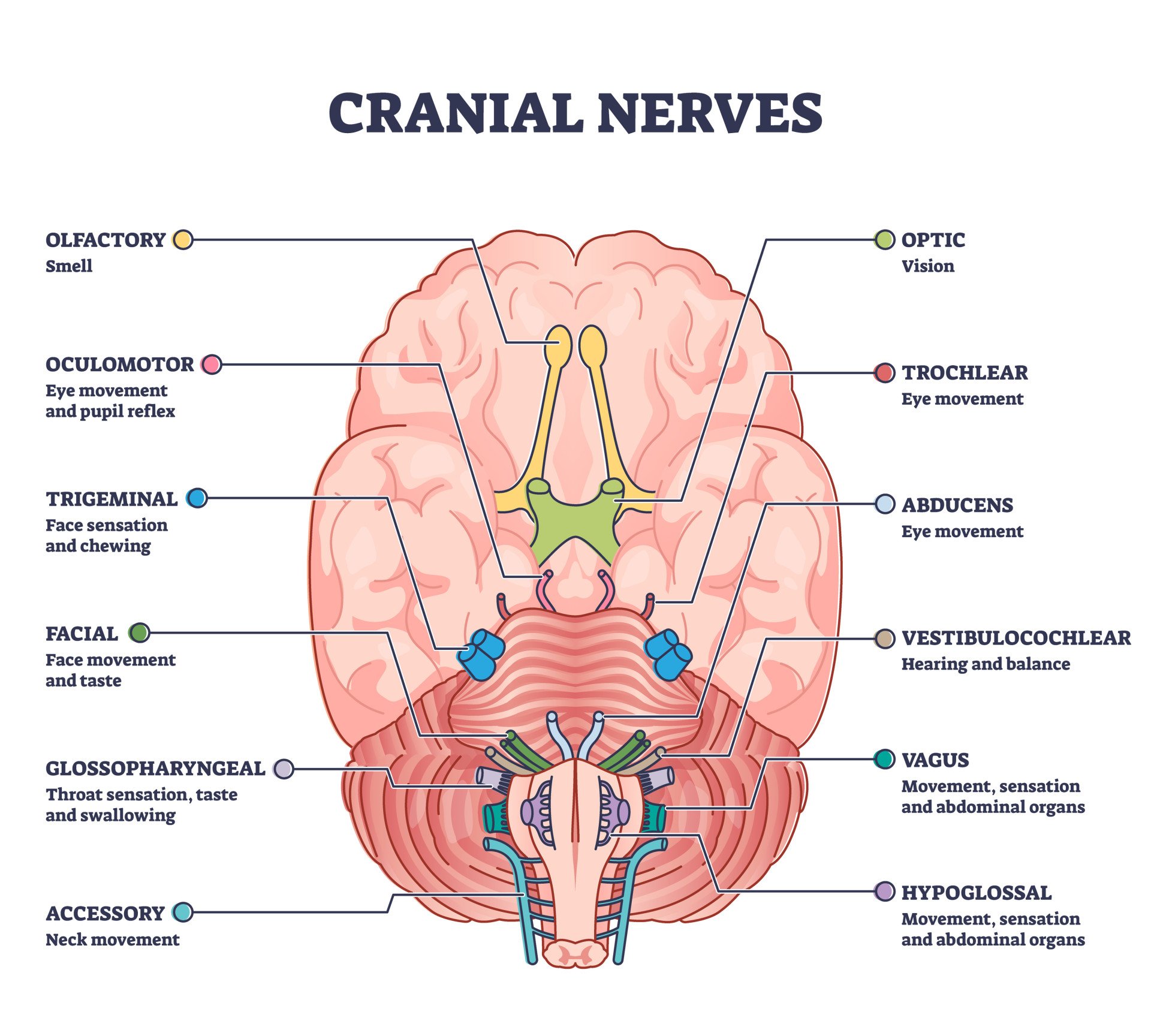
Inside the cranium (the dome of the skull), there are 12 nerves, called cranial nerves:
- Cranial nerve 1: The first is the olfactory nerve, which allows for your sense of smell.
- Cranial nerve 2: The optic nerve governs eyesight.
- Cranial nerve 3: The oculomotor nerve controls pupil response and other motions of the eye, and branches out from the area in the brainstem where the midbrain meets the pons.
- Cranial nerve 4: The trochlear nerve controls muscles in the eye. It emerges from the back of the midbrain part of the brainstem.
- Cranial nerve 5: The trigeminal nerve is the largest and most complex of the cranial nerves, with both sensory and motor function. It originates from the pons and conveys sensation from the scalp, teeth, jaw, sinuses, parts of the mouth and face to the brain, allows the function of chewing muscles, and much more.
- Cranial nerve 6: The abducens nerve innervates some of the muscles in the eye.
- Cranial nerve 7: The facial nerve supports face movement, taste, glandular and other functions.
- Cranial nerve 8: The vestibulocochlear nerve facilitates balance and hearing.
- Cranial nerve 9: The glossopharyngeal nerve allows taste, ear and throat movement, and has many more functions.
- Cranial nerve 10: The vagus nerve allows sensation around the ear and the digestive system and controls motor activity in the heart, throat and digestive system.
- Cranial nerve 11: The accessory nerve innervates specific muscles in the head, neck and shoulder.
- Cranial nerve 12: The hypoglossal nerve supplies motor activity to the tongue.
The first two nerves originate in the cerebrum, and the remaining 10 cranial nerves emerge from the brainstem, which has three parts: the midbrain, the pons and the medulla.
Find a Treatment Center
- Neurology and Neurosurgery
Find Additional Treatment Centers at:
- Howard County Medical Center
- Sibley Memorial Hospital
- Suburban Hospital

Request an Appointment

Moyamoya Disease

Motor Stereotypies

Related Topics
- Brain, Nerves and Spine
- Alzheimer's disease & dementia
- Arthritis & Rheumatism
- Attention deficit disorders
- Autism spectrum disorders
- Biomedical technology
- Diseases, Conditions, Syndromes
- Endocrinology & Metabolism
- Gastroenterology
- Gerontology & Geriatrics
- Health informatics
- Inflammatory disorders
- Medical economics
- Medical research
- Medications
- Neuroscience
- Obstetrics & gynaecology
- Oncology & Cancer
- Ophthalmology
- Overweight & Obesity
- Parkinson's & Movement disorders
- Psychology & Psychiatry
- Radiology & Imaging
- Sleep disorders
- Sports medicine & Kinesiology
- Vaccination
- Breast cancer
- Cardiovascular disease
- Chronic obstructive pulmonary disease
- Colon cancer
- Coronary artery disease
- Heart attack
- Heart disease
- High blood pressure
- Kidney disease
- Lung cancer
- Multiple sclerosis
- Myocardial infarction
- Ovarian cancer
- Post traumatic stress disorder
- Rheumatoid arthritis
- Schizophrenia
- Skin cancer
- Type 2 diabetes
- Full List »
share this!
September 9, 2021
How the brain solves problems
by Delia Du Toit, Wits University
In trying to think of an introduction for this article it occurred to me that had I been inside an MRI, the screen would have showed several brain regions lighting up like Times Square as my mind was attempting to solve the problem.
First, the prefrontal cortex, basal ganglia and thalamus would recognize that the blank page meant that there was a problem that needed to be solved. The thought that the editor might not favor this first-person account in a science article would send the limbic system , the primal part of the brain where emotions are processed, into overdrive. The amygdala, that little almond-shaped nugget at the base of the brain, would look like a Christmas tree as anxiety ticked up.
Finally, as words started filling the screen, the prefrontal cortex behind the forehead would flicker and flash. The hippocampus would access memories of previous similar articles, the information-gathering process and even school-level English classes decades ago, to help the process along. And all this activity would happen at once.
Holistic problem solving
Depending on the problem in front of you, the entire brain could be involved in trying to find a solution, says Professor Kate Cockcroft, Division Leader of cognitive neuroscience at the Neuroscience Research Laboratory (Wits NeuRL) in the School of Human and Community Development.
"You would use many different brain regions to solve a problem, especially a novel or difficult one. The idea of processes being localized in one or two parts of the brain has been replaced with newer evidence that it is the connections among brain areas and their interaction that is important in cognitive processes . Some areas may be more activated with certain problems—a visual problem would activate the visual cortices, for example.
"All this activity takes place as electrochemical signals. The signals form within neurons, pass along the branch-like axons and jump from one neuron to the next across gaps called synapses, with the help of neurotransmitter chemicals. The pattern, size, shape and number of these signals, what they communicate with, and the region of the brain in which they happen, determine what they achieve."
Although problem solving is a metacognitive—"thinking about thinking"—process, that does not make it solely the domain of the highly evolved human prefrontal cortex , adds Dr. Sahba Besharati, Division Leader of social-affective neuroscience at NeuRL.
"This is the most recently evolved part of the human brain, but problem solving does not happen in isolation—it's immersed in a social context that influences how we interpret information. Your background, gender, religion or emotions, among other factors, all influence how you interpret a problem. This means that it would involve other brain areas like the limbic system, one of the oldest brain systems housed deep within the cortex," says Besharati.
"Problem-solving abilities are not a human peculiarity. Some animals are even better than us at solving certain problems, but we all share basic problem-solving skills—if there's danger, leave; if you're hungry, find food."
None of this would be possible without memory either, says Cockcroft. "Without it, we would forget what it is that we are trying to solve and we wouldn't be able to use past experiences to help us solve it."
And memory is, again, linked to emotion. "We use this information to increase the likelihood of positive results when solving new problems," she says.
Improving your skills
It has been proven time and again that just about any brain process can be improved—including problem-solving abilities. "Brain plasticity is a real thing—the brain can reorganize itself with targeted intervention," says Besharati. "Rehabilitation from neurological injury is a dynamic process and an ever-improving science that has allowed us to understand how the brain can change and adapt in response to the environment. Studies have also shown that simple memorisation exercises can assist tremendously in retaining cognitive skills in old age."
Of course, all these processes depend on your brain recognizing that there's a problem to be dealt with in the first place—if you don't realize you're spending money foolishly, you can't improve your finances. "Recognition of a problem can happen at both a conscious and unconscious level. Stroke patients who are not aware of their motor paralysis, for example, deludedly don't believe that they are paralyzed and will sometimes not engage in rehabilitation. But their delusions often spontaneously recover, suggesting recognition at an unconscious level and that, over time, the brain can restore function."
If all else fails, there might be some value to the adage "sleep on it," says Cockcroft. "Sleep is believed to assist memory consolidation—changing memories from a fragile state in which they can easily be damaged to a permanent state. In doing so, they become stored in different brain regions and new neural connections are formed that may assist problem solving. On waking, you may have formed associations between information that you didn't think of previously. This seems to be most effective within three hours of learning new information—perhaps we should institute compulsory naps for students after lectures!"
Explore further
Feedback to editors

Study identifies first drug therapy for sleep apnea
16 hours ago

Research suggests potential targets for prevention and early identification of psychotic disorders
18 hours ago

C. elegans study finds mRNA balance in cells influences lifespan

Mapping the heart to prevent damage caused by a heart attack

Study reveals evolution of human cold and menthol sensing protein, offers hope for future non-addictive pain therapies

Study uncovers hidden DNA mechanisms of rare genetic diseases
19 hours ago

Popular diabetes drugs may reduce the risk of dementia

Using digital technology and data to sustain intermittent fasting and improve health outcomes: One man's journey
20 hours ago

Study finds connection between cannabis use and increased risk of severe COVID-19

Activating a molecular target reverses multiple hallmarks of aging, new study demonstrates
Related stories.

Have a vexing problem? Sleep on it.
Oct 17, 2019

Can't solve a riddle? The answer might lie in knowing what doesn't work
Mar 4, 2021

Researchers question the existence of the social brain as a separate system
Oct 15, 2020

Creativity important to lift math education
Feb 4, 2020

A child's brain activity reveals their memory ability
May 25, 2020

Why people with depression can sometimes experience memory problems
Feb 9, 2021
Recommended for you

Brain health is rooted in state of mind, finds study
23 hours ago

Newly discovered subtypes and sex differences give insight into molecular mechanisms of ALS
21 hours ago

Clinical trial reports promising new treatment reduces suffering in Sanfilippo syndrome

Scans show brain's estrogen activity changes during menopause
22 hours ago

Research reveals how sighted and blind people's brains change when they learn to echolocate

Imaging technology captures how neurons communicate with new clarity
Let us know if there is a problem with our content.
Use this form if you have come across a typo, inaccuracy or would like to send an edit request for the content on this page. For general inquiries, please use our contact form . For general feedback, use the public comments section below (please adhere to guidelines ).
Please select the most appropriate category to facilitate processing of your request
Thank you for taking time to provide your feedback to the editors.
Your feedback is important to us. However, we do not guarantee individual replies due to the high volume of messages.
E-mail the story
Your email address is used only to let the recipient know who sent the email. Neither your address nor the recipient's address will be used for any other purpose. The information you enter will appear in your e-mail message and is not retained by Medical Xpress in any form.
Newsletter sign up
Get weekly and/or daily updates delivered to your inbox. You can unsubscribe at any time and we'll never share your details to third parties.
More information Privacy policy
Donate and enjoy an ad-free experience
We keep our content available to everyone. Consider supporting Science X's mission by getting a premium account.
E-mail newsletter
Frontal Lobe: What It Is, Function, Location & Damage
Olivia Guy-Evans, MSc
Associate Editor for Simply Psychology
BSc (Hons) Psychology, MSc Psychology of Education
Olivia Guy-Evans is a writer and associate editor for Simply Psychology. She has previously worked in healthcare and educational sectors.
Learn about our Editorial Process
Saul Mcleod, PhD
Editor-in-Chief for Simply Psychology
BSc (Hons) Psychology, MRes, PhD, University of Manchester
Saul Mcleod, PhD., is a qualified psychology teacher with over 18 years of experience in further and higher education. He has been published in peer-reviewed journals, including the Journal of Clinical Psychology.
On This Page:
The frontal lobe is the brain’s largest region, located behind the forehead, at the front of the brain. These lobes are part of the cerebral cortex and are the largest brain structure.
The frontal lobe’s main functions are typically associated with ‘higher’ cognitive functions, including decision-making, problem-solving, thought, and attention .
It contains the motor cortex , which is involved in planning and coordinating movement; the prefrontal cortex, which is responsible for higher-level cognitive functioning; and Broca’s Area , which is essential for language production.

Frontal Lobe Functions
Below is a list of some of the associated functions of the frontal lobe:
Executive processes (capacity to plan, organize, initiate, and self-monitor) Voluntary behavior Problem-solving Voluntary motor control Intelligence Language processing Language comprehension Self-control Emotional control
The frontal lobes are believed to be our behavior and emotional control centers, meaning that this area is activated when needing to control our behaviors to be socially appropriate and to control our emotional responses, especially in social situations.
Moreover, the frontal lobes are thought to be the home of our personalities.
Alike to most lobes in the brain, there are two frontal lobes located in the left and right hemispheres.
Each lobe controls the operations on opposite sides of the body: the left hemisphere controls the right side of the body and vice versa.
It is believed the left frontal lobe is the most dominant lobe and works predominantly with language, logical thinking, and analytical reasoning.
The right frontal lobe, on the other hand, is most associated with non-verbal abilities, creativity, imagination, and musical and art skills.
The frontal lobe, like other structures of the brain, does not always work in isolation from each other. The frontal lobes work alongside other brain regions in order to control a variety of functions.
Substructures
The frontal lobe contains the motor cortex , which is involved in planning and coordinating movement; the prefrontal cortex, which is responsible for higher-level cognitive functioning; and Broca’s area, which is essential for language production.
Prefrontal Cortex
The prefrontal cortex is primarily responsible for the ‘higher’ brain functions of the frontal lobes, including decision-making, problem-solving, intelligence, and emotion regulation.
This area has also been found to be associated with the social skills and personality of humans.
This idea is supported by the famous case study of Phineas Gage , whose personality changed after losing a part of his prefrontal cortex after an iron rod impaled his head.
The frontal cortex has also been shown to be activated when an experience becomes conscious. Different ideas and perceptions are bound together in this region, both of which are necessary for conscious experience. Concluding that this area may be especially important for consciousness.
Cognitive disorders that have been shown to be linked to this region are attention deficit hyperactivity disorder ( ADHD ), Autism, bipolar disorder, depression , and schizophrenia.
The prefrontal cortex can be further divided into the dorsolateral prefrontal cortex and the orbitofrontal cortex.
Motor and Premotor Cortex
The motor cortex is critical for initiating motor movements, as well as coordinating motor movements, hence why it is called the motor cortex.
Each area of the motor cortex corresponds precisely with specific body parts. For instance, there is an area that controls the left and the right foot.
The premotor cortex is associated with planning and executing motor movements. Within this area, voluntary movement is rehearsed, distinguishing these movements from unconscious reactions.
The premotor cortex has also been shown to be important for imitation learning through the use of mirror neurons. These neurons essentially allow us to reflect the body language, facial expressions, and emotions of others.
Furthermore, the prefrontal cortex can support cognitive functions of a social kind, such as showing empathy.
Broca’s Area
Another region of the frontal lobes worth mentioning is Broca’s area . This region is located in the dominant hemisphere of the frontal lobes, which is the left side for around 97% of humans.
This region is associated with the production of speech and written language, as well as with the processing and comprehension of language.
The name is taken from the French scientist Paul Broca, whose work with language-impaired patients led him to conclude that we speak with our left brains.
Language differences in those with Autism may be correlated to differences in the structure and function of Broca’s area (Bauman & Kemper, 2005).
As the frontal lobes are situated at the front of the brain and are large in size, this makes them more susceptible to damage. This area is the most common for traumatic brain injuries, with damage to this region causing a variety of symptoms.
Below is a list of symptoms that may occur if an individual has experienced damage within their frontal lobe:
- Changes in mood
- Attention deficits
- Atypical social skills
- Difficulty problem-solving
- Lack of impulse control/ risk-taking
- Loss of spontaneity in social interactions
- Reduced motivation
- Impaired judgment
- Reduced creativity
Damage to Broca’s area, in particular, has been shown to affect the ability to speak, understand language, and produce coherent sentences.
One of the most famous case studies associated with frontal lobe damage is the case of Phineas Gage . He was a railway construction worker who suffered an unfortunate accident when a metal rod impaled his brain in the frontal region.
Gage survived this accident but was said to have experienced some personality changes because of the trauma. Before the accident, Gage was described as a ‘well-balanced’ and smart, energetic person.
After his accident, he was described as being childlike in his intellectual capacities and had a loss of social inhibition (behaving in ways that were considered socially inappropriate).
This case study implies that the frontal lobes are essential to our personalities, intelligence, and social skills. As well as trauma to the head is a cause of damage to the frontal lobes, there are many other causes that can lead to damage.
For instance, a brain tumor, stroke, or infection can cause deficits in this lobe. Similarly, conditions such as cerebral palsy, Huntington’s disease, dementia, or other neurodegenerative diseases can lead to associated damage.
If someone is suspected of having frontal lobe damage, there are methods to diagnose this. Magnetic resonance imaging (MRI) and computerized tomography (CT) scans can detect some differences in the frontal lobes after suffering a stroke or infection, as well as be able to detect dementia.
Also, neuropsychological evaluations can be completed to test for areas such as speech comprehension, social behavior, memory, problem-solving, and impulse control, among others.
One common test to establish frontal lobe damage is the Wisconsin Card Sorting task. Within this task, individuals will be shown cards of varying sorts, such as some having symbols, numbers, different shapes, and colors on them.
They will then be asked to sort the cards by a certain criterion, which will then change throughout the test. Those who have damage to a certain part of the frontal lobes may struggle with this task and will not adjust to new sorting criteria. They will stick with the original criteria (this is called perseveration).
Other tests worth noting as finger tapping tests, to test for motor skill ability, and the Token Test, which tests for language skills.
To be able to treat frontal lobe damage, occupational, speech, and physical therapy can be helpful for rehabilitating these lost or damaged skills.
Finally, a talking therapy called cognitive behavioral therapy (CBT) is common for working on regulating emotions and aiding impulse behaviors.
CBT may not fully treat physical damage to the frontal lobes, but it can help those with impairments cope and manage their symptoms.
Research Studies
- Semmes, Weinstein, Ghent & Teuber (1963) suggested that the frontal lobes played a part in spatial orientation, particularly our body’s orientation in space.
- Eslinger & Grattan (1993) investigated damage to the prefrontal cortex. They suggested that people with damage to this area may not have problems with word comprehension or identifying objects by their names, but if asked to say or write as many words as possible or describe as many uses of an object, they would find this task difficult. This shows that damage to one area associated with language does not impair all aspects of language.
- Kolb & Milner (1981) discussed the involvement of the frontal lobes in facial expressions. They found that patients with frontal lobe damage had difficulty expressing spontaneous facial expressions and would also show fewer facial movements spontaneously.
- Kaufman, Geyer & Milstein (2017) reported that patients who suffered damage to their frontal lobes had changes to their personalities.
- It was found that these patients developed an abrupt, suspicious, and sometimes even argumentative manner.
- Some patients were reported to have displayed ‘emotional incontinence,’ whereby they would have bouts of pathological laughing or crying.
- Walker & Blummer (1975) found that damage to the frontal lobes resulted in displays of abnormal sexual behavior in the orbital region and reduced sexual interest if the dorsolateral region was damaged.
- Stuss et al. (1992) found that damage to certain areas of the frontal lobes resulted in ‘bland’ personalities. These patients also displayed fewer signs of distress in emotionally heightened situations.
- Catani et al. (2016) investigated the brains of people with Autism and found support for the hypothesis that Autism is associated with different connectivity in the frontal lobe region compared with neurotypical individuals.
- Mubarik & Tohid (2016) conducted a literature review of studies that investigated the frontal lobes of those with schizophrenia.
- They found that many people with schizophrenia have differences in the structure of white matter , grey matter , and functional activity in their frontal lobes compared to those without the condition.
Bauman, M. L., & Kemper, T. L. (2005). Neuroanatomic observations of the brain in autism: a review and future directions. International Journal of Developmental Neuroscience, 23 (2-3), 183-187.
Catani, M., Dell’Acqua, F., Budisavljevic, S., Howells, H., Thiebaut de Schotten, M., Froudist-Walsh, S., … & Murphy, D. G. (2016). Frontal networks in adults with autism spectrum disorder. Brain, 139 (2), 616-630.
Eslinger, P. J., & Grattan, L. M. (1993). Frontal lobe and frontal-striatal substrates for different forms of human cognitive flexibility. Neuropsychologia, 31 (1), 17-28.
Kaufman, D. M., Geyer, H. L., & Milstein, M. J. (2017). Chapter 21-neurotransmitters and drug abuse. Kaufman’s Clinical Neurology for Psychiatrists . 8th ed. Amsterdam: Elsevier, 495-517.
Kolb, B., & Milner, B. (1981). Performance of complex arm and facial movements after focal brain lesions. Neuropsychologia, 19( 4), 491-503.
Mubarik, A., & Tohid, H. (2016). Frontal lobe alterations in schizophrenia: a review. Trends in Psychiatry and Psychotherapy , 38(4), 198-206.
Semmes, J., Weinstein, S., GHENT, G., Meyer, J. S., & Teuber, H. L. (1963). Correlates of impaired orientation in personal and extrapersonal space. Brain, 86 (4), 747-772.
Stuss, D. T., Ely, P., Hugenholtz, H., Richard, M. T., LaRochelle, S., Poirier, C. A., & Bell, I. (1985). Subtle neuropsychological deficits in patients with good recovery after closed head injury. Neurosurgery, 17 (1), 41-47.
Walker, A. E., & Blumer, D. (1975). The localization of sex in the brain. In Cerebral localization (pp. 184-199). Springer, Berlin, Heidelberg.

Related Articles
Biopsychology
Summary of the Cranial Nerves
Parts of the Brain: Anatomy, Structure & Functions
Amygdala: What It Is & Its Functions
Autonomic Nervous System (ANS): What It Is and How It Works
Biopsychology , Psychology
Biological Approach In Psychology
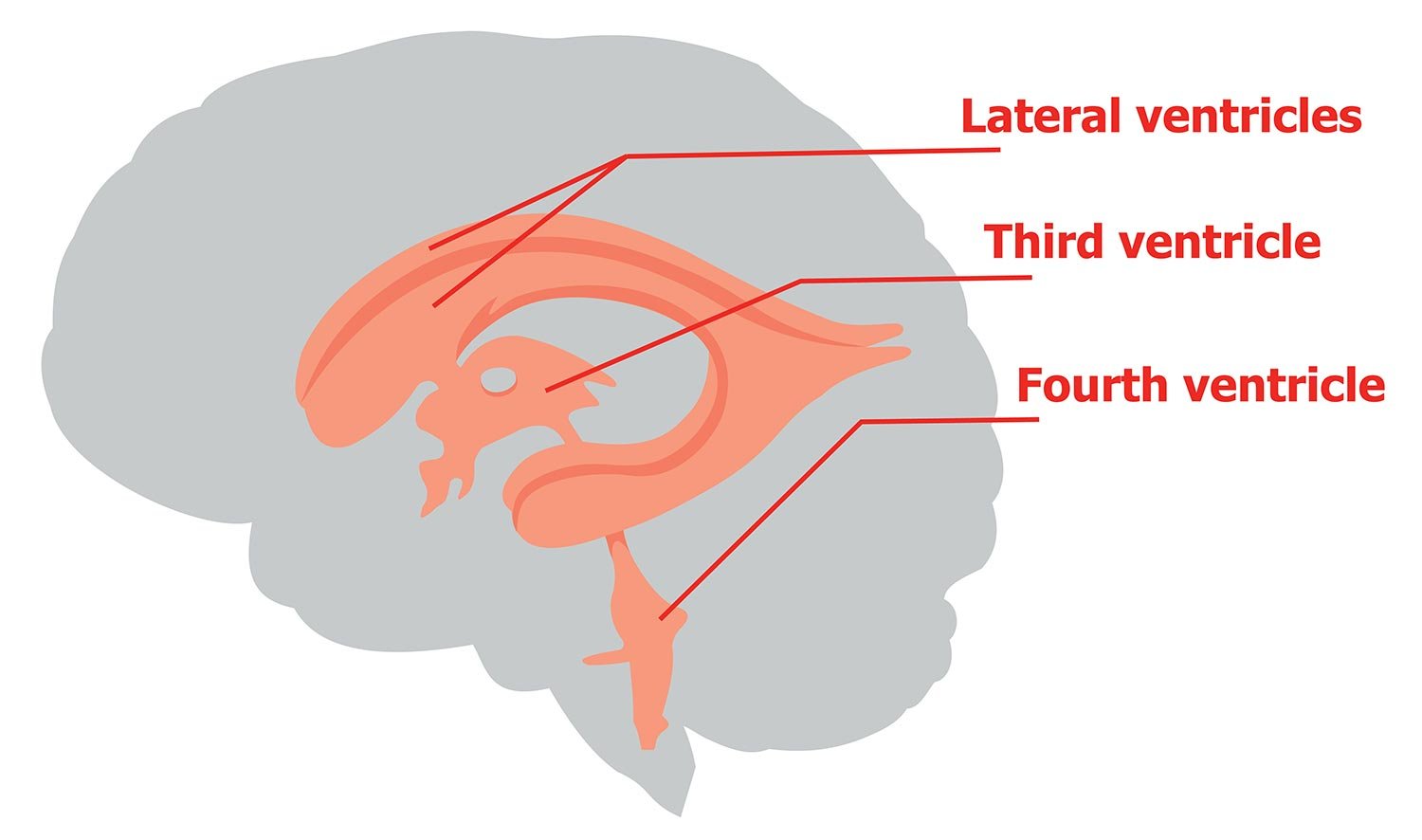
Ventricles of the Brain
- Brain Development
- Childhood & Adolescence
- Diet & Lifestyle
- Emotions, Stress & Anxiety
- Learning & Memory
- Thinking & Awareness
- Alzheimer's & Dementia
- Childhood Disorders
- Immune System Disorders
- Mental Health
- Neurodegenerative Disorders
- Infectious Disease
- Neurological Disorders A-Z
- Body Systems
- Cells & Circuits
- Genes & Molecules
- The Arts & the Brain
- Law, Economics & Ethics
- Neuroscience in the News
- Supporting Research
- Tech & the Brain
- Animals in Research
- BRAIN Initiative
- Meet the Researcher
- Neuro-technologies
- Tools & Techniques
Core Concepts
- For Educators
- Ask an Expert
- The Brain Facts Book

Important Brain Regions for Decision-Making
- Reviewed 8 May 2023
- Author Marissa Fessenden
- Source BrainFacts/SfN

You make many different types of decisions every day. Some of these rely primarily on logical reasoning — for example, when you compare the timetables for the bus and subway to determine the quickest way to get to a friend’s house. Other decisions have emotional consequences at stake, like when the person you’re trying to impress offers you a cigarette — your desire to be accepted might outweigh your rational consideration of smoking’s harms.
Decision-making requires a person to weigh values, understand rules, make plans, and form predictions about the outcomes of their choices. Both logical reasoning and emotional (affective) decision-making involve the brain’s prefrontal cortex (PFC). In particular, activity in the lateral PFC is especially important in overriding emotional responses during decision-making. The area’s strong connections with brain regions related to motivation and emotion, such as the amygdala and nucleus accumbens , seem to exert a sort of top-down control over emotional and impulsive responses. For example, brain imaging studies have found the lateral PFC is more active in people declining a small monetary reward given immediately in favor of receiving a larger reward in the future. This is one of the last areas of the brain to mature — usually in a person’s late 20s — which explains why teens sometimes may have trouble regulating emotions or controlling impulses.
The orbitofrontal cortex , a region of the PFC located just behind the eyes, also appears to be important in affective decision-making, especially in situations involving reward and punishment. The area has been implicated in addiction as well as social behavior.
Adapted from the 8th edition of Brain Facts by Marissa Fessenden.
About the Author

Marissa Fessenden
Marissa Fessenden is a freelance science journalist and illustrator. They gravitate toward stories about genes, wildlife and places large and small, as well as times when art gets science-y or science gets artsy.
CONTENT PROVIDED BY
BrainFacts/SfN
What to Read Next

Also In Thinking & Awareness

Popular articles on BrainFacts.org

BrainFacts Book
Download a copy of the newest edition of the book, Brain Facts: A Primer on the Brain and Nervous System.
Ask An Expert
Ask a neuroscientist your questions about the brain.
Submit a Question
A beginner's guide to the brain and nervous system.

SUPPORTING PARTNERS
- Privacy Policy
- Accessibility Policy
- Terms and Conditions
- Manage Cookies
Some pages on this website provide links that require Adobe Reader to view.
- Bipolar Disorder
- Therapy Center
- When To See a Therapist
- Types of Therapy
- Best Online Therapy
- Best Couples Therapy
- Best Family Therapy
- Managing Stress
- Sleep and Dreaming
- Understanding Emotions
- Self-Improvement
- Healthy Relationships
- Student Resources
- Personality Types
- Guided Meditations
- Verywell Mind Insights
- 2024 Verywell Mind 25
- Mental Health in the Classroom
- Editorial Process
- Meet Our Review Board
- Crisis Support
Overview of the Problem-Solving Mental Process
Kendra Cherry, MS, is a psychosocial rehabilitation specialist, psychology educator, and author of the "Everything Psychology Book."
:max_bytes(150000):strip_icc():format(webp)/IMG_9791-89504ab694d54b66bbd72cb84ffb860e.jpg)
Rachel Goldman, PhD FTOS, is a licensed psychologist, clinical assistant professor, speaker, wellness expert specializing in eating behaviors, stress management, and health behavior change.
:max_bytes(150000):strip_icc():format(webp)/Rachel-Goldman-1000-a42451caacb6423abecbe6b74e628042.jpg)
- Identify the Problem
- Define the Problem
- Form a Strategy
- Organize Information
- Allocate Resources
- Monitor Progress
- Evaluate the Results
Frequently Asked Questions
Problem-solving is a mental process that involves discovering, analyzing, and solving problems. The ultimate goal of problem-solving is to overcome obstacles and find a solution that best resolves the issue.
The best strategy for solving a problem depends largely on the unique situation. In some cases, people are better off learning everything they can about the issue and then using factual knowledge to come up with a solution. In other instances, creativity and insight are the best options.
It is not necessary to follow problem-solving steps sequentially, It is common to skip steps or even go back through steps multiple times until the desired solution is reached.
In order to correctly solve a problem, it is often important to follow a series of steps. Researchers sometimes refer to this as the problem-solving cycle. While this cycle is portrayed sequentially, people rarely follow a rigid series of steps to find a solution.
The following steps include developing strategies and organizing knowledge.
1. Identifying the Problem
While it may seem like an obvious step, identifying the problem is not always as simple as it sounds. In some cases, people might mistakenly identify the wrong source of a problem, which will make attempts to solve it inefficient or even useless.
Some strategies that you might use to figure out the source of a problem include :
- Asking questions about the problem
- Breaking the problem down into smaller pieces
- Looking at the problem from different perspectives
- Conducting research to figure out what relationships exist between different variables
2. Defining the Problem
After the problem has been identified, it is important to fully define the problem so that it can be solved. You can define a problem by operationally defining each aspect of the problem and setting goals for what aspects of the problem you will address
At this point, you should focus on figuring out which aspects of the problems are facts and which are opinions. State the problem clearly and identify the scope of the solution.
3. Forming a Strategy
After the problem has been identified, it is time to start brainstorming potential solutions. This step usually involves generating as many ideas as possible without judging their quality. Once several possibilities have been generated, they can be evaluated and narrowed down.
The next step is to develop a strategy to solve the problem. The approach used will vary depending upon the situation and the individual's unique preferences. Common problem-solving strategies include heuristics and algorithms.
- Heuristics are mental shortcuts that are often based on solutions that have worked in the past. They can work well if the problem is similar to something you have encountered before and are often the best choice if you need a fast solution.
- Algorithms are step-by-step strategies that are guaranteed to produce a correct result. While this approach is great for accuracy, it can also consume time and resources.
Heuristics are often best used when time is of the essence, while algorithms are a better choice when a decision needs to be as accurate as possible.
4. Organizing Information
Before coming up with a solution, you need to first organize the available information. What do you know about the problem? What do you not know? The more information that is available the better prepared you will be to come up with an accurate solution.
When approaching a problem, it is important to make sure that you have all the data you need. Making a decision without adequate information can lead to biased or inaccurate results.
5. Allocating Resources
Of course, we don't always have unlimited money, time, and other resources to solve a problem. Before you begin to solve a problem, you need to determine how high priority it is.
If it is an important problem, it is probably worth allocating more resources to solving it. If, however, it is a fairly unimportant problem, then you do not want to spend too much of your available resources on coming up with a solution.
At this stage, it is important to consider all of the factors that might affect the problem at hand. This includes looking at the available resources, deadlines that need to be met, and any possible risks involved in each solution. After careful evaluation, a decision can be made about which solution to pursue.
6. Monitoring Progress
After selecting a problem-solving strategy, it is time to put the plan into action and see if it works. This step might involve trying out different solutions to see which one is the most effective.
It is also important to monitor the situation after implementing a solution to ensure that the problem has been solved and that no new problems have arisen as a result of the proposed solution.
Effective problem-solvers tend to monitor their progress as they work towards a solution. If they are not making good progress toward reaching their goal, they will reevaluate their approach or look for new strategies .
7. Evaluating the Results
After a solution has been reached, it is important to evaluate the results to determine if it is the best possible solution to the problem. This evaluation might be immediate, such as checking the results of a math problem to ensure the answer is correct, or it can be delayed, such as evaluating the success of a therapy program after several months of treatment.
Once a problem has been solved, it is important to take some time to reflect on the process that was used and evaluate the results. This will help you to improve your problem-solving skills and become more efficient at solving future problems.

A Word From Verywell
It is important to remember that there are many different problem-solving processes with different steps, and this is just one example. Problem-solving in real-world situations requires a great deal of resourcefulness, flexibility, resilience, and continuous interaction with the environment.
Get Advice From The Verywell Mind Podcast
Hosted by therapist Amy Morin, LCSW, this episode of The Verywell Mind Podcast shares how you can stop dwelling in a negative mindset.
Follow Now : Apple Podcasts / Spotify / Google Podcasts
You can become a better problem solving by:
- Practicing brainstorming and coming up with multiple potential solutions to problems
- Being open-minded and considering all possible options before making a decision
- Breaking down problems into smaller, more manageable pieces
- Asking for help when needed
- Researching different problem-solving techniques and trying out new ones
- Learning from mistakes and using them as opportunities to grow
It's important to communicate openly and honestly with your partner about what's going on. Try to see things from their perspective as well as your own. Work together to find a resolution that works for both of you. Be willing to compromise and accept that there may not be a perfect solution.
Take breaks if things are getting too heated, and come back to the problem when you feel calm and collected. Don't try to fix every problem on your own—consider asking a therapist or counselor for help and insight.
If you've tried everything and there doesn't seem to be a way to fix the problem, you may have to learn to accept it. This can be difficult, but try to focus on the positive aspects of your life and remember that every situation is temporary. Don't dwell on what's going wrong—instead, think about what's going right. Find support by talking to friends or family. Seek professional help if you're having trouble coping.
Davidson JE, Sternberg RJ, editors. The Psychology of Problem Solving . Cambridge University Press; 2003. doi:10.1017/CBO9780511615771
Sarathy V. Real world problem-solving . Front Hum Neurosci . 2018;12:261. Published 2018 Jun 26. doi:10.3389/fnhum.2018.00261
By Kendra Cherry, MSEd Kendra Cherry, MS, is a psychosocial rehabilitation specialist, psychology educator, and author of the "Everything Psychology Book."
- Type 2 Diabetes
- Heart Disease
- Digestive Health
- Multiple Sclerosis
- Diet & Nutrition
- Supplements
- Health Insurance
- Public Health
- Patient Rights
- Caregivers & Loved Ones
- End of Life Concerns
- Health News
- Thyroid Test Analyzer
- Doctor Discussion Guides
- Hemoglobin A1c Test Analyzer
- Lipid Test Analyzer
- Complete Blood Count (CBC) Analyzer
- What to Buy
- Editorial Process
- Meet Our Medical Expert Board
The Anatomy of the Prefrontal Cortex
Associated conditions, frequently asked questions.
The prefrontal cortex is an important part of your brain. It is at the front of the frontal lobe, which is immediately behind the forehead. It affects your behavior, personality, and ability to plan. This article will explain more about the anatomy, location, and function of the prefrontal cortex.
Eric Raptosh Photography / Getty Images
The prefrontal cortex (PFC) is connected to many other parts of the brain and is able to send and receive information. The prefrontal cortex is divided into these two parts:
- Medial PFC (mPFC) : It is involved in self-reflection, memory, and emotional processing.
- Lateral PFC (lPFC) : It is involved in sensory processing, motor control, and performance monitoring.
The prefrontal cortex is involved in many brain functions. One of the most important is executive function , or the ability to self-regulate and plan ahead. Examples of executive function include:
- Controlling your behavior and impulses
- Delaying instant gratification
- Regulating your emotions
- Making decisions
- Solving problems
- Making long-term goals
- Balancing short-term rewards with future goals
- Changing your behavior when situations change
- Seeing and predicting the consequences of your behavior
- Being able to consider many streams of information
- Being able to focus your attention
The prefrontal cortex also affects your personality. A historical example of what happens when a person’s prefrontal cortex is damaged occurred in the mid-1800s. When railroad worker Phineas Gage’s prefrontal cortex was damaged by a metal rod going through his skull, he survived, but his personality changed. He became impulsive and lost the ability to plan.
Damage to the prefrontal cortex can happen from:
- Brain trauma : Accidents, falls, sports injuries, and physical altercations can cause a traumatic brain injury.
- Cancer : Cancer originating in the brain (primary tumors) or spreading to the brain from other original sites ( metastatic brain tumors ) can cause damage.
- Tumors : In addition to cancerous tumors, benign (noncancerous) brain tumors can harm the prefrontal cortex.
- Stroke : A blocked blood vessel or bleed in the brain can damage the prefrontal cortex.
When the prefrontal cortex is damaged, it may cause the following conditions:
- Changes in personality and behavior
- Problems with social behavior and an increase in antisocial behavior
- Higher chance of committing violence or stealing
- Problems regulating emotions and impulses
- Attention deficit hyperactivity disorder (ADHD) : A neurodevelopmental condition that affects attention, hyperactivity, and impulsiveness
- Post-traumatic stress disorder (PTSD) : A mental health disorder that affects people after traumatic events
- Schizophrenia : A mental health condition that affects a person’s behavior, thoughts, and feelings
- Bipolar disorder : A condition that causes extreme mood swings
If you have damage to the prefrontal cortex or another condition that is affecting it, your healthcare provider may start with a physical exam and a mental status exam. These tests will help them evaluate your thinking and rule out other conditions.
To check your brain, a healthcare provider may order the following tests:
- Magnetic resonance imaging (MRI) : Detailed images taken using magnetic fields
- Computed tomography (CT) scan : A detailed computerized X-ray scan
- Positron-emission tomography (PET scan) : Imaging that uses a radioactive tracer to look for cells that are active
The prefrontal cortex is found in front of the frontal lobe of the brain. It affects your behavior, personality, and executive function. When the prefrontal cortex is damaged, it can cause changes to how you think and behave.
A Word From Verywell
It is important to remember that you may not always notice changes in your behavior or thinking. Your friends and loved ones are more likely to point out that something is wrong.
Even if you think everything is fine, it is worth having a conversation with your healthcare provider and checking on your brain health. It is better to catch problems earlier and get treatment.
Yes, the prefrontal cortex grows as a person matures from childhood to early adulthood. It is one of the last parts of the brain to develop completely.
In general, the prefrontal cortex is considered fully developed by the age of 25. This is why car insurance companies charge higher rates until a person turns 25 because they are high-risk drivers. They explain that the prefrontal cortex is involved in risk-taking and decision-making, which are both important for driving.
A person may lose executive function if the prefrontal cortex is damaged. They may still be able to do some tasks and even work, but they will have trouble planning and controlling their behavior.
Yes, it is possible for a person to survive without a prefrontal cortex. However, not having this portion of the brain will have an enormous impact on their ability to reason, plan ahead, control behavior, and solve problems. A person’s ability to have social relationships would also be affected severely.
Grossmann T. The role of medial prefrontal cortex in early social cognition . Front Hum Neurosci . 2013;7:340. doi:10.3389/fnhum.2013.00340
Arain M, Haque M, Johal L, et al. Maturation of the adolescent brain . Neuropsychiatr Dis Treat . 2013;9:449-461. doi:10.2147/NDT.S39776
Teles RV. Phineas Gage's great legacy . Dement Neuropsychol . 2020;14(4):419-421. doi:10.1590/1980-57642020dn14-040013
Barrash J, Bruss J, Anderson SW, et al. Lesions in different prefrontal sectors are associated with different types of acquired personality disturbances . Cortex. 2022;147:169-184. doi:10.1016/j.cortex.2021.12.004
By Lana Bandoim Bandoim has nearly 20 years of experience writing for a variety of outlets including health sites, scientific publishers, and academic medical centers.
- Share full article
Advertisement
Supported by
What Your Brain Looks Like When It Solves a Math Problem

By Benedict Carey
- July 28, 2016
Solving a hairy math problem might send a shudder of exultation along your spinal cord. But scientists have historically struggled to deconstruct the exact mental alchemy that occurs when the brain successfully leaps the gap from “Say what?” to “Aha!”
Now, using an innovative combination of brain-imaging analyses, researchers have captured four fleeting stages of creative thinking in math. In a paper published in Psychological Science, a team led by John R. Anderson, a professor of psychology and computer science at Carnegie Mellon University, demonstrated a method for reconstructing how the brain moves from understanding a problem to solving it, including the time the brain spends in each stage.
The imaging analysis found four stages in all: encoding (downloading), planning (strategizing), solving (performing the math), and responding (typing out an answer).
“I’m very happy with the way the study worked out, and I think this precision is about the limit of what we can do” with the brain imaging tools available, said Dr. Anderson, who wrote the report with Aryn A. Pyke and Jon M. Fincham, both also at Carnegie Mellon.
To capture these quicksilver mental operations, the team first taught 80 men and women how to interpret a set of math symbols and equations they had not seen before. The underlying math itself wasn’t difficult, mostly addition and subtraction, but manipulating the newly learned symbols required some thinking. The research team could vary the problems to burden specific stages of the thinking process — some were hard to encode, for instance, while others extended the length of the planning stage.
The scientists used two techniques of M.R.I. data analysis to sort through what the participants’ brains were doing. One technique tracked the neural firing patterns during the solving of each problem; the other identified significant shifts from one kind of mental state to another. The subjects solved 88 problems each, and the research team analyzed the imaging data from those solved successfully.
The analysis found four separate stages that, depending on the problem, varied in length by a second or more. For instance, planning took up more time than the other stages when a clever workaround was required. The same stages are likely applicable to solving many creative problems, not just in math. But knowing how they play out in the brain should help in designing curriculums, especially in mathematics, the paper suggests.
“We didn’t know exactly what students were doing when they solved problems,” said Dr. Anderson, whose lab designs math instruction software. “Having a clearer understanding of that will help us develop better instruction; I think that’s the first place this work will have some impact.”
Like the Science Times page on Facebook. | Sign up for the Science Times newsletter.

In order to continue enjoying our site, we ask that you confirm your identity as a human. Thank you very much for your cooperation.

- News/Events
- Arts and Sciences
- Design and the Arts
- Engineering
- Global Futures
- Health Solutions
- Nursing and Health Innovation
- Public Service and Community Solutions
- University College
- Thunderbird School of Global Management
- Polytechnic
- Downtown Phoenix
- Online and Extended
- Lake Havasu
- Research Park
- Washington D.C.
- Biology Bits
- Bird Finder
- Coloring Pages
Experiments and Activities
- Games and Simulations
- Quizzes in Other Languages
- Virtual Reality (VR)
- World of Biology
- Meet Our Biologists
- Listen and Watch
- PLOSable Biology
- All About Autism
- Xs and Ys: How Our Sex Is Decided
- When Blood Types Shouldn’t Mix: Rh and Pregnancy
- What Is the Menstrual Cycle?
- Understanding Intersex
- The Mysterious Case of the Missing Periods
- Summarizing Sex Traits
- Shedding Light on Endometriosis
- Periods: What Should You Expect?
- Menstruation Matters
- Investigating In Vitro Fertilization
- Introducing the IUD
- How Fast Do Embryos Grow?
- Helpful Sex Hormones
- Getting to Know the Germ Layers
- Gender versus Biological Sex: What’s the Difference?
- Gender Identities and Expression
- Focusing on Female Infertility
- Fetal Alcohol Syndrome and Pregnancy
- Ectopic Pregnancy: An Unexpected Path
- Creating Chimeras
- Confronting Human Chimerism
- Cells, Frozen in Time
- EvMed Edits
- Stories in Other Languages
- Virtual Reality
- Zoom Gallery
- Ugly Bug Galleries
- Ask a Question
- Top Questions
- Question Guidelines
- Permissions
- Information Collected
- Author and Artist Notes
- Share Ask A Biologist
- Articles & News
- Our Volunteers
- Teacher Toolbox

show/hide words to know
Disorder: something that is not in order. Not arranged correctly. In medicine a disorder is when something in the body is not working correctly.
Electroencephalogram: visual recording showing the electrical activity of the brain (EEG)... more
Emotion: any of a long list of feelings a person can have such as joy, anger and love... more
What Are the Regions of the Brain and What Do They Do?
The brain has many different parts . The brain also has specific areas that do certain types of work. These areas are called lobes. One lobe works with your eyes when watching a movie. There is a lobe that is controlling your legs and arms when running and kicking a soccer ball. There are two lobes that are involved with reading and writing. Your memories of a favorite event are kept by the same lobe that helps you on a math test. The brain is controlling all of these things and a lot more. Use the map below to take a tour of the regions in the brain and learn what they control in your body.
The brain is a very busy organ. It is the control center for the body. It runs your organs such as your heart and lungs. It is also busy working with other parts of your body. All of your senses – sight, smell, hearing, touch, and taste – depend on your brain. Tasting food with the sensors on your tongue is only possible if the signals from your taste buds are sent to the brain. Once in the brain, the signals are decoded. The sweet flavor of an orange is only sweet if the brain tells you it is.
| | |
Brain Waves
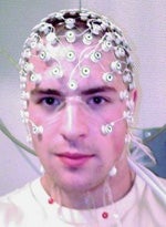
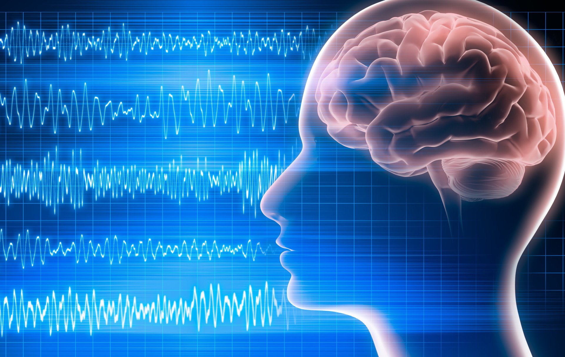
How do you tell if the brain is working? What is it doing and how do you measure it? The head gear on the right that looks like it's from a work of science fiction measures electrical activity in the brain. These electrical waves are called brain waves.
When neurons send a signal they use electrical currents to pass messages to other nearby neurons. Just one or two neurons signaling is too small a change to be noticed. When a huge group of neurons signal at once, however, they can be recorded and measured with the help of special tools.
Measuring electrical activity in the brain is usually done with electrodes. Electrodes are devices able to record electrical changes over time. These are attached to the surface of the skin in specific places around the head. Recordings of brain wave activity look like a series of waves. These are called electroencephalograms, or EEGs for short.
Measuring activity in the brain can be a very useful tool in scientific studies. They can also be used to help identify sleeping disorders and other medical conditions relating to the brain.

The first human electroencephalogram, recorded in 1924 by Hans Berger.
Computer animation credit: BodyParts3D, Copyright© 2010 The Database Center for Life Science licensed under CC Attribution-Share Alike 2.1 Japan.
Read more about: A Nervous Journey
View citation, bibliographic details:.
- Article: What's Your Brain Doing?
- Author(s): Brett Szymik
- Publisher: Arizona State University School of Life Sciences Ask A Biologist
- Site name: ASU - Ask A Biologist
- Date published: May 9, 2011
- Date accessed: June 12, 2024
- Link: https://askabiologist.asu.edu/brain-regions
Brett Szymik. (2011, May 09). What's Your Brain Doing?. ASU - Ask A Biologist. Retrieved June 12, 2024 from https://askabiologist.asu.edu/brain-regions
Chicago Manual of Style
Brett Szymik. "What's Your Brain Doing?". ASU - Ask A Biologist. 09 May, 2011. https://askabiologist.asu.edu/brain-regions
MLA 2017 Style
Brett Szymik. "What's Your Brain Doing?". ASU - Ask A Biologist. 09 May 2011. ASU - Ask A Biologist, Web. 12 Jun 2024. https://askabiologist.asu.edu/brain-regions
Computer animation image of the human brain. The colors show the frontal lobe (red), parietal lobe (orange), temporal lobe (green), and occipital lobe (yellow).
A Nervous Journey
Coloring Pages and Worksheets
Neuron Anatomy
What's In Your Brain
What's Your Brain Doing?

Be Part of Ask A Biologist
By volunteering, or simply sending us feedback on the site. Scientists, teachers, writers, illustrators, and translators are all important to the program. If you are interested in helping with the website we have a Volunteers page to get the process started.
Share to Google Classroom

How the Brain Combines Memories to Solve Problems
Summary: Using AI technology, researchers provide new insight into how the human brain connects individual episodic memories to help solve problems.
Source: Cell Press.
Humans have the ability to creatively combine their memories to solve problems and draw new insights, a process that depends on memories for specific events known as episodic memory. But although episodic memory has been extensively studied in the past, current theories do not easily explain how people can use their episodic memories to arrive at these novel insights.
Results from a team of neuroscientists and artificial intelligence researchers at DeepMind, Otto von Guericke University Magdeburg and the German Center for Neurodegenerative Diseases (DZNE), publishing in the journal Neuron on September 19, provide a window into the way the human brain connects individual episodic memories to solve problems.
For example, imagine you see a woman driving a car on your street. The next day, you see a man driving the exact same car on your street. This might trigger the memory of the woman you saw the day before, and you might reason that the pair live together, given that they share a car.
The researchers propose a novel brain mechanism that would allow retrieved memories to trigger the retrieval of further, related memories in this way. This mechanism allows the retrieval of multiple linked memories, which then enable the brain to create new kinds of insights like these.
In common with standard theories of episodic memory, the authors posit that individual memories are stored as separate memory traces in a brain region called the hippocampus.
“Episodic memories can tell you whether you have met someone before, or where you parked your car,” says Raphael Koster (@raphael_koster), a researcher at DeepMind (@DeepMindAI). “The hippocampal system supports this type of memory, which is crucial for rapid learning.”
Unlike standard theories, the new theory explores a neglected anatomical connection that loops out of the hippocampus to the neighboring entorhinal cortex but then immediately passes back in. It is this recurrent connection, the researchers thought, that allows memories retrieved from the hippocampus to trigger the retrieval of further, related memories.
The researchers devised a way of testing this theory by taking high-resolution 7-Tesla functional MRI scans from 26 young men and women as they performed a task that required them to draw insights across separate events.
The volunteers were shown pairs of photographs: one of a face and one of an object or a place. Each individual object and place appeared in two separate photo pairs, each of which included a different face. This meant that every photo pair was linked with another pair through the shared object or place image.
In a second phase of the experiment, the researchers tested whether the participants could infer the indirect connection between these linked pairs of photos by showing one face and asking the participants to choose between two other faces. One of the choices–the correct one–had been paired with the same object or place image, and one had not.
The researchers guessed that the presented face would trigger the retrieval of the paired object or place and thus spark brain activity that would pass out of the hippocampus into the entorhinal cortex. Crucially, the researchers also expected to see evidence that this activity would then pass back into the hippocampus to trigger the retrieval of the correct linked face.
“Using specialized techniques developed in our lab in Magdeburg, we were able to separate out the parts of the entorhinal cortex that provide the input to the hippocampus,” says Yi Chen, researcher at Otto von Guericke University. “This allowed us to precisely measure the patterns of activation in the hippocampus input and output separately.”
The researchers trained a computer algorithm to distinguish between activation for scenes and objects within these input and output regions. The algorithm was then applied when only faces were displayed on the screen. If the algorithm indicated the presence of scene or object information on these trials, it could only be driven by retrieved memories of the linked scene or object photos.
“Our data showed that when the hippocampus retrieves a memory, it doesn’t just pass it to the rest of the brain,” says DeepMind’s Dharshan Kumaran (@dharshsky). “Instead, it recirculates the activation back into the hippocampus, triggering the retrieval of other related memories.”
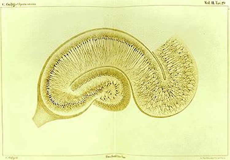
The researchers think of the algorithm’s results as a synthesis of new and old theories.
“The results could be thought of as the best of both worlds: you preserve the ability to remember individual experiences by keeping them separate, while at the same time allowing related memories to be combined on the fly at the point of retrieval,” says Kumaran. “This ability is useful for understanding how the different parts of a story fit together, for example–something not possible if you just retrieve a single memory.”
The authors believe that their results could help AI learn faster in the future.
“While there are many domains where AI is superior, humans still have an advantage when tasks depend on the flexible use of episodic memory,” says Martin Chadwick (@MartinJChadwick), another researcher at DeepMind. “If we can understand the mechanisms that allow people to do this, the hope is that we can replicate them within our AI systems, providing them with a much greater capacity for rapidly solving novel problems.”
Funding: This research was funded by DeepMind and the German Research foundation.
Source: Erin Kohnke – Cell Press Publisher: Organized by NeuroscienceNews.com. Image Source: NeuroscienceNews.com image is in the public domain. Original Research: Open access research for “Big-Loop Recurrence within the Hippocampal System Supports Integration of Information across Episodes” by Raphael Koster, Martin J. Chadwick, Yi Chen, David Berron, Andrea Banino, Emrah Düzel, Demis Hassabis, and Dharshan Kumaran in Neuron . Published September 19 2018. doi: 10.1016/j.neuron.2018.08.009
[cbtabs][cbtab title=”MLA”]Cell Press”How the Brain Combines Memories to Solve Problems.” NeuroscienceNews. NeuroscienceNews, 19 September 2018. <https://neurosciencenews.com/memory-problem-solving-9891/>.[/cbtab][cbtab title=”APA”]Cell Press(2018, September 19). How the Brain Combines Memories to Solve Problems. NeuroscienceNews . Retrieved September 19, 2018 from https://neurosciencenews.com/memory-problem-solving-9891/[/cbtab][cbtab title=”Chicago”]Cell Press”How the Brain Combines Memories to Solve Problems.” https://neurosciencenews.com/memory-problem-solving-9891/ (accessed September 19, 2018).[/cbtab][/cbtabs]
Big-Loop Recurrence within the Hippocampal System Supports Integration of Information across Episodes
Recent evidence challenges the widely held view that the hippocampus is specialized for episodic memory, by demonstrating that it also underpins the integration of information across experiences. Contemporary computational theories propose that these two contrasting functions can be accomplished by big-loop recurrence, whereby the output of the system is recirculated back into the hippocampus. We use ultra-high-resolution fMRI to provide support for this hypothesis, by showing that retrieved information is presented as a new input on the superficial entorhinal cortex—driven by functional connectivity between the deep and superficial entorhinal layers. Further, the magnitude of this laminar connectivity correlated with inferential performance, demonstrating its importance for behavior. Our findings offer a novel perspective on information processing within the hippocampus and support a unifying framework in which the hippocampus captures higher-order structure across experiences, by creating a dynamic memory space from separate episodic codes for individual experiences.
Love to read about developments in the interests of all people. Wish that scientists took a firmer stance against the militarization of our lives.
Comments are closed.
ALS Study Reveals Subtypes and Promising Drug Target

Cannabis Use Linked to Severe COVID-19
Ozempic May Reduce Alzheimer’s Risk
Resilience Linked to Healthier Brain and Gut
- Published on May 29, 2023
- May 29, 2023
What Part of the Brain Controls Thinking? Here’s How It Affects You

Jump to section

With more than 86 billion functional neurons, the brain is the most complex organ in the human body that deals with thinking. It controls everything it does and thinks.
In other words, it’s the “boss of your body.” So, many people wonder, “What part of the brain controls thinking?”
The thing is, this particular organ plays such a crucial role in our whole system. It develops functions for the five senses: sight, sound, touch, taste, and hearing. And it helps primary functions such as breathing, talking, storing memories, and thinking.
It’s our most precious gift, according to Jim Kwik , international brain coach and trainer of Mindvalley’s Superbrain Quest. Why? Simply because it’s what “ allows us to learn, love, think, create, and even to experience joy. ”
Not only that, it’s “ the gateway to our emotions, to our capacity for deeply experiencing life, to our ability to have lasting intimacy ,” as well as helping us innovate, grow, and accomplish.
Which Part of the Brain Controls Thinking?
The brain consists of three main parts:
- The cerebrum. It’s the outer part of the brain, which consists of the frontal, parietal, temporal, and occipital lobes.
- The cerebellum. It’s located at the bottom of the brain, near the back of your head.
- The brain stem. This third part is located beneath the cerebrum and in front of the cerebellum in the brain stem.

These three parts control processes in the body, including movement, memory, and thinking.
The role of the cerebrum
The cerebrum makes up more than 85% of the brain’s weight. It’s the part of the brain that controls daily activities such as reading, learning, and speech. It also assists planned muscle movements such as walking, running, and body movement.
The cerebrum is the thinking part of the brain. It helps you play chess, solve a crossword puzzle, or figure out your next move in a complex video game.
The cerebrum has two hemispheres—the left hemisphere and the right hemisphere. Each hemisphere controls the opposite side of the body.
The two hemispheres have four sections called lobes—frontal, parietal, occipital, and temporal. Each of these lobes controls specific aspects of the thinking process.
The role of the cerebellum
If the brain is a super-efficient, bustling office complex, the cerebellum is the office’s facilities manager, maintaining the heartbeat of operations.
It works behind the scenes, taking care of fine motor movements, balance, and coordination. You know, the day-to-day tasks, like making sure you can navigate through your house without bumping into furniture or helping you catch a football mid-air on a Sunday afternoon.
Although it isn’t directly involved in thinking, the cerebellum plays an important role in this process. This part of the brain takes up to 10% of its total volume yet contains more than half of all the neurons in the brain.
Scientists have discovered that the “unconscious” cerebellum interacts with the “conscious” cerebrum to perform functions. The cerebellum carries out planned muscle movements such as running and jumping. That’s why sometimes scientists call it the “thinking cerebellum.”
Research nowadays suggests that the cerebellum is a key player in predicting our emotional reactions. The study found that “the cerebellum, which was initially considered to be mainly involved in motor coordination and execution, is now recognized as an associative center for higher cognitive and emotional functions even in the developing brain.”
The brainstem
Tucked away at the base of our brain is the brainstem. It connects the brain to the spinal cord and holds the reins of many automatic, essential bodily functions.
Here’s one way to look at it: Imagine your body as a busy city. The brainstem, then, would be akin to its efficient and diligent city hall, managing essential services like heart rate, breathing, and blood pressure.
These are our body’s critical life-support systems, running constantly in the background, much like a city’s water and power supply.
The brainstem also acts as a vital communication highway, ferrying messages between the brain and the rest of the body. It’s a bit like the city’s central post office, handling the constant flow of information to and from various parts of the body.
Within its compact structure, the brainstem houses numerous nuclei involved in different functions and relaying sensory information. It’s also where cranial nerves originate, controlling functions from eye movement to facial sensations and movements.
Other Brain Regions You Should Know About
Different brain activities are linked to different parts of the brain. Here are a few of the main ones explained:
Which part of the brain controls critical thinking?
When it comes to which part of the brain controls critical thinking and intelligence, we have to consider that the prefrontal cortex, anterior cingulate cortex, and parietal lobe work together like a highly trained Olympic relay team.
Each player has its own role: the prefrontal cortex in decision-making, the anterior cingulate cortex in conflict detection, and the parietal lobe in information processing. Understanding this synchrony can unlock our true critical thinking potential.
Which part of the brain controls memory?
When it comes to the art of memory, we turn our attention to the hippocampus.
Nestled deep within the brain, this small region takes on the monumental task of memory formation and recall. It stores information of past experiences and opens up the space to create new ones.
Learn more: What Part of the Brain Controls Memory?
Which part of the brain controls breathing?
Breathe in, breathe out—a simple act that sustains life and is meticulously regulated by the medulla oblongata in our brainstem.
This unsung hero of our nervous system ensures the continuity of this essential function, similar to the ceaseless rhythm of the ocean tides. Acknowledging its role can help us appreciate the fascinating intricacies of our bodies.
Learn more: What Part of the Brain Controls Breathing?
Which part of the brain deals with emotions?
Now, you know which parts of the brain control thinking and memory. But what about our emotions?
All positive and negative emotions and spontaneous feelings, from excitement to sadness, are processed in the limbic system. This system controls your emotions and interacts with other parts of the brain.
At the same time, another part of the brain called the amygdala handles emotional reactions such as love, hate, and sexual desire.
Learn more: The Anatomy of Feelings: What Part of the Brain Controls Emotions?

Unleash Your Superbrain
With centuries of research, the human brain remains the biggest mystery in the world. It is the most complex part of the body and controls movement, sight, and thinking. And of course, it’s the part of our system that’s most closely related to our minds.
And as Jim says, “We need to understand how our minds work so we can work our minds better.”
If you need some guidance to start unlocking your superbrain and mind, Mindvalley is the place to be. With transformational quests such as Superbrain , guided by Jim Kwik, you’ll master your mind’s functions in no time. And you can embark on a journey of:
- Techniques for supercharging your memory, focus, and learning capacity
- The most beneficial brain diet
- How to clear your mind of unwanted thoughts
- How to transform the way your brain is wired
- Achieving top performance when learning
By unlocking your free access , you can sample classes from this program and many others. All you have to do is open your mind to your greatest potential. And don’t be afraid to take the first step.
Welcome in.
Recommended Free Masterclass For You

Discover Powerful Hacks to Unlock Your Superbrain to Learn Faster, Comprehend More and Forget Less
Join the foremost expert in memory improvement and brain performance, Jim Kwik, in a free masterclass that will dive into the one skill you will ever need — learning how to learn Reserve My Free Spot Now
Alexandra Tudor
Jim Kwik is the trainer of Mindvalley’s Superbrain and Super Reading Quests. He’s a brain coach and a world expert in speed reading, memory improvement, and optimal brain performance. Known as the “boy with the broken brain” due to a childhood injury, Jim discovered strategies to dramatically enhance his mental performance. He is now committed to helping people improve their memory, learn to speed-read, increase their decision-making skills, and turn on their superbrain. He has shared his techniques with Hollywood actors, Fortune 500 companies, and trailblazing entrepreneurs like Elon Musk and Richard Branson to reach their highest level of mental performance. He is also one of the most sought-after trainers for top organizations like Harvard University, Nike, Virgin, and GE.
How we reviewed this article
2020 study: the cerebellum’s role in movement and cognition, consensus paper: the cerebellum’s role in movement and cognition, you might also like.

Get Started
- Try Mindvalley for Free
- Free Masterclasses
- Coaching Certifications
- Vishen Lakhiani
- The Mindvalley Show
- Partnerships
- In English 🇺🇸
- En Español 🇪🇸
- © 2024 Mindvalley, Inc.
- English (EN)
Fact-Checking: Our Process
Mindvalley is committed to providing reliable and trustworthy content.
We rely heavily on evidence-based sources, including peer-reviewed studies and insights from recognized experts in various personal growth fields. Our goal is to keep the information we share both current and factual.
The Mindvalley fact-checking guidelines are based on:
- Content Foundation: Our articles build upon Mindvalley’s quest content, which are meticulously crafted and vetted by industry experts to ensure foundational credibility and reliability.
- Research and Sources: Our team delves into credible research, ensuring every piece is grounded in facts and evidence, offering a holistic view on personal growth topics.
- Continuous Updates: In the dynamic landscape of personal development, we are committed to keeping our content fresh. We often revisit and update our resources to stay abreast of the latest developments.
- External Contributions: We welcome insights from external contributors who share our passion for personal transformation and consciousness elevation.
- Product Recommendations and Affiliations: Recommendations come after thoughtful consideration and alignment with Mindvalley’s ethos, grounded in ethical choices.
To learn more about our dedication to reliable reporting, you can read our detailed editorial standards .

February 1, 2024
18 min read
Brains Are Not Required When It Comes to Thinking and Solving Problems—Simple Cells Can Do It
Tiny clumps of cells show basic cognitive abilities, and some animals can remember things after losing their head
By Rowan Jacobsen
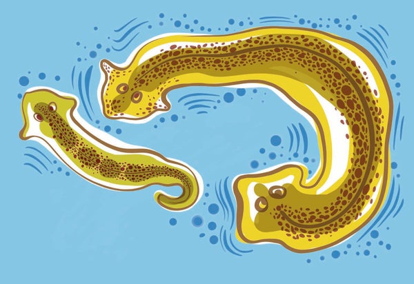
Natalya Balnova
T he planarian is nobody's idea of a genius. A flatworm shaped like a comma, it can be found wriggling through the muck of lakes and ponds worldwide. Its pin-size head has a microscopic structure that passes for a brain. Its two eyespots are set close together in a way that makes it look cartoonishly confused. It aspires to nothing more than life as a bottom-feeder.
But the worm has mastered one task that has eluded humanity's greatest minds: perfect regeneration. Tear it in half, and its head will grow a new tail while its tail grows a new head. After a week two healthy worms swim away.
Growing a new head is a neat trick. But it's the tail end of the worm that intrigues Tufts University biologist Michael Levin. He studies the way bodies develop from single cells , among other things, and his research led him to suspect that the intelligence of living things lies outside their brains to a surprising degree. Substantial smarts may be in the cells of a worm's rear end, for instance. “All intelligence is really collective intelligence, because every cognitive system is made of some kind of parts,” Levin says. An animal that can survive the complete loss of its head was Levin's perfect test subject.
On supporting science journalism
If you're enjoying this article, consider supporting our award-winning journalism by subscribing . By purchasing a subscription you are helping to ensure the future of impactful stories about the discoveries and ideas shaping our world today.
In their natural state planaria prefer the smooth and sheltered to the rough and open. Put them in a dish with a corrugated bottom, and they will huddle against the rim. But in his laboratory, about a decade ago, Levin trained some planaria to expect yummy bits of liver puree that he dripped into the middle of a ridged dish. They soon lost all fear of the rough patch, eagerly crossing the divide to get the treats. He trained other worms in the same way but in smooth dishes. Then he decapitated them all.
Levin discarded the head ends and waited two weeks while the tail ends regrew new heads. Next he placed the regenerated worms in corrugated dishes and dripped liver into the center. Worms that had lived in a smooth dish in their previous incarnation were reluctant to move. But worms regenerated from tails that had lived in rough dishes learned to go for the food more quickly. Somehow, despite the total loss of their brains, those planaria had retained the memory of the liver reward. But how? Where?
It turns out that regular cells—not just highly specialized brain cells such as neurons—have the ability to store information and act on it. Now Levin has shown that the cells do so by using subtle changes in electric fields as a type of memory. These revelations have put the biologist at the vanguard of a new field called basal cognition. Researchers in this burgeoning area have spotted hallmarks of intelligence—learning, memory, problem-solving—outside brains as well as within them.
Until recently, most scientists held that true cognition arrived with the first brains half a billion years ago. Without intricate clusters of neurons, behavior was merely a kind of reflex. But Levin and several other researchers believe otherwise. He doesn't deny that brains are awesome, paragons of computational speed and power. But he sees the differences between cell clumps and brains as ones of degree, not kind. In fact, Levin suspects that cognition probably evolved as cells started to collaborate to carry out the incredibly difficult task of building complex organisms and then got souped-up into brains to allow animals to move and think faster.
That position is being embraced by researchers in a variety of disciplines, including roboticists such as Josh Bongard, a frequent Levin collaborator who runs the Morphology, Evolution, and Cognition Laboratory at the University of Vermont. “Brains were one of the most recent inventions of Mother Nature, the thing that came last,” says Bongard, who hopes to build deeply intelligent machines from the bottom up. “It's clear that the body matters, and then somehow you add neural cognition on top. It's the cherry on the sundae. It's not the sundae.”
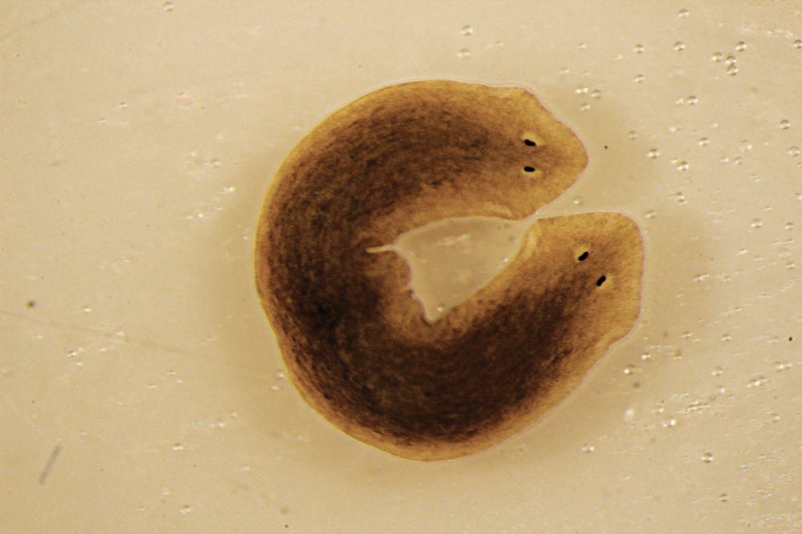
Head cells in the flatworm Dugesia japonica have different bioelectric voltages than tail cells do. Switch the voltages around and cut off the tail, and the head will regenerate a second head. Credit: Michael Levin
In recent years interest in basal cognition has exploded as researchers have recognized example after example of surprisingly sophisticated intelligence at work across life's kingdoms, no brain required. For artificial-intelligence scientists such as Bongard, basal cognition offers an escape from the trap of assuming that future intelligences must mimic the brain-centric human model. For medical specialists, there are tantalizing hints of ways to awaken cells' innate powers of healing and regeneration.
And for the philosophically minded, basal cognition casts the world in a sparkling new light. Maybe thinking builds from a simple start. Maybe it is happening all around us, every day, in forms we haven't recognized because we didn't know what to look for. Maybe minds are everywhere.
Although it now seems like a Dark Ages idea, only a few decades ago many scientists believed that nonhuman animals couldn't experience pain or other emotions. Real thought? Out of the question. The mind was the purview of humans. “It was the last beachhead,” says Pamela Lyon of the University of Adelaide, a scholar of basal cognition, who coined the term for the field in 2018. Lyon sees scientists' insistence that human intelligence is qualitatively different as just another doomed form of exceptionalism. “We've been ripped from every central position we've inhabited,” she points out. Earth is not the center of the universe. People are just another animal species. But real cognition—that was supposed to set us apart.
Now that notion, too, is in retreat as researchers document the rich inner lives of creatures increasingly distant from us. Apes, dogs, dolphins, crows and even insects are proving more savvy than suspected. In his 2022 book The Mind of a Bee , behavioral ecologist Lars Chittka chronicles his decades of work with honeybees, showing that bees can use sign language, recognize individual human faces, and remember and convey the locations of far-flung flowers . They have good moods and bad, and they can be traumatized by near-death experiences such as being grabbed by an animatronic spider hidden in a flower. (Who wouldn't be?)
But bees, of course, are animals with actual brains, so a soupçon of smarts doesn't really shake the paradigm. The bigger challenge comes from evidence of surprisingly sophisticated behavior in our brainless relatives. “The neuron is not a miracle cell,” says Stefano Mancuso, a University of Florence botanist who has written several books on plant intelligence. “It's a normal cell that is able to produce an electric signal. In plants almost every cell is able to do that.”
On one plant, the touch-me-not, feathery leaves normally fold and wilt when touched (a defense mechanism against being eaten), but when a team of scientists at the University of Western Australia and the University of Firenze in Italy conditioned the plant by jostling it throughout the day without harming it, it quickly learned to ignore the stimulus. Most remarkably, when the scientists left the plant alone for a month and then retested it, it remembered the experience. Other plants have other abilities. A Venus flytrap can count, snapping shut only if two of the sensory hairs on its trap are tripped in quick succession and pouring digestive juices into the closed trap only if its sensory hairs are tripped three more times.
These responses in plants are mediated by electric signals, just as they are in animals. Wire a flytrap to a touch-me-not, and you can make the entire touch-me-not collapse by touching a sensory hair on the flytrap. And these and other plants can be knocked out by anesthetic gas. Their electric activity flatlines, and they stop responding as if unconscious.
Plants can sense their surroundings surprisingly well. They know whether they are being shaded by part of themselves or by something else. They can detect the sound of running water (and will grow toward it) and of bees' wings (and will produce nectar in preparation). They know when they are being eaten by bugs and will produce nasty defense chemicals in response. They even know when their neighbors are under attack: when scientists played a recording of munching caterpillars to a cress plant, that was enough for the plant to send a surge of mustard oil into its leaves.
Plants' most remarkable behavior tends to get underappreciated because we see it every day: they seem to know exactly what form they have and plan their future growth based on the sights, sounds and smells around them, making complicated decisions about where future resources and dangers might be located in ways that can't be boiled down to simple formulas. As Paco Calvo, director of the Minimal Intelligence Laboratory at the University of Murcia in Spain and author of Planta Sapiens , puts it, “Plants have to plan ahead to achieve goals, and to do so, they need to integrate vast pools of data. They need to engage with their surroundings adaptively and proactively, and they need to think about the future. They just couldn't afford to do otherwise.”
None of this implies that plants are geniuses, but within their limited tool set, they show a solid ability to perceive their world and use that information to get what they need—key components of intelligence. But again, plants are a relatively easy case—no brains but lots of complexity and trillions of cells to play with. That's not the situation for single-celled organisms, which have traditionally been relegated to the “mindless” category by virtually everyone. If amoebas can think, then humans need to rethink all kinds of assumptions.
Yet the evidence for cogitating pond scum grows daily. Consider the slime mold, a cellular puddle that looks a bit like melted Velveeta and oozes through the world's forests digesting dead plant matter. Although it can be the size of a throw rug, a slime mold is one single cell with many nuclei. It has no nervous system, yet it is an excellent problem solver. When researchers from Japan and Hungary placed a slime mold at one end of a maze and a pile of oat flakes at the other, the slime mold did what slime molds do, exploring every possible option for tasty resources. But once it found the oat flakes, it retreated from all the dead ends and concentrated its body in the path that led to the oats, choosing the shortest route through the maze (of four possible solutions) every time. Inspired by that experiment, the same researchers then piled oat flakes around a slime mold in positions and quantities meant to represent the population structure of Tokyo, and the slime mold contorted itself into a very passable map of the Tokyo subway system.
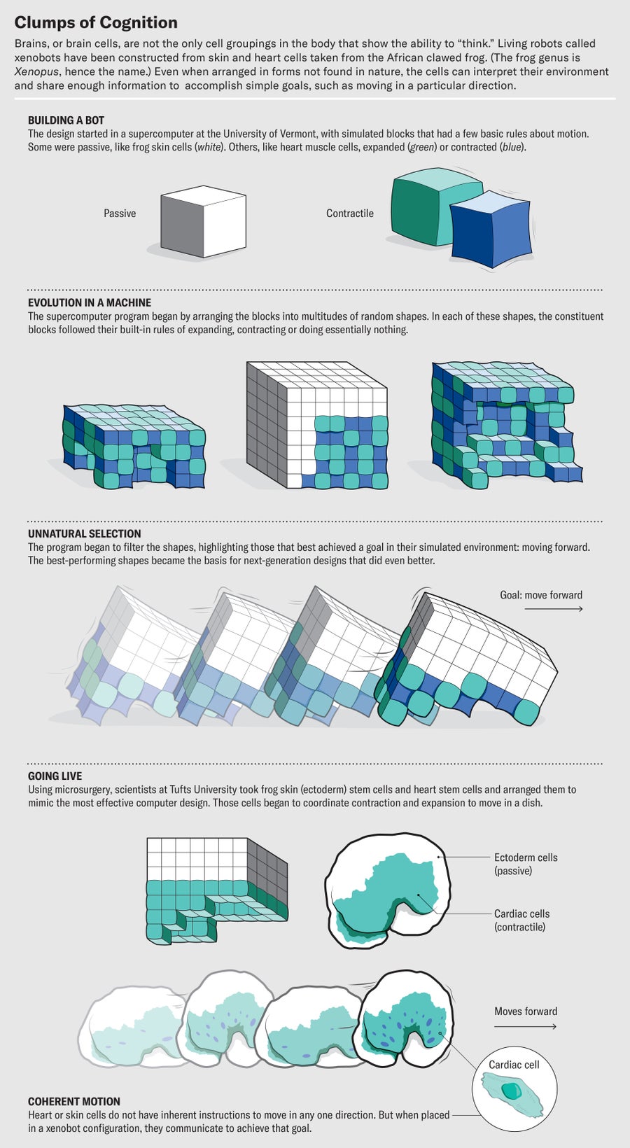
Credit: Brown Bird Design; Source: “A Scalable Pipeline for Designing Reconfigurable Organisms,” by Sam Kriegman et al., in PNAS , Vol. 117; January 2020 ( reference )
Such problem-solving could be dismissed as simple algorithms, but other experiments make it clear that slime molds can learn. When Audrey Dussutour of France's National Center for Scientific Research placed dishes of oatmeal on the far end of a bridge lined with caffeine (which slime molds find disgusting), slime molds were stymied for days, searching for a way across the bridge like an arachnophobe trying to scooch past a tarantula. Eventually they got so hungry that they went for it, crossing over the caffeine and feasting on the delicious oatmeal, and soon they lost all aversion to the formerly distasteful stuff. They had overcome their inhibitions and learned from the experience, and they retained the memory even after being put into a state of suspended animation for a year.
Which brings us back to the decapitated planaria. How can something without a brain remember anything? Where is the memory stored? Where is its mind?
T he orthodox view of memory is that it is stored as a stable network of synaptic connections among neurons in a brain. “That view is clearly cracking,” Levin says. Some of the demolition work has come from the lab of neuroscientist David Glanzman of the University of California, Los Angeles. Glanzman was able to transfer a memory of an electric shock from one sea slug to another by extracting RNA from the brains of shocked slugs and injecting it into the brains of new slugs. The recipients then “remembered” to recoil from the touch that preceded the shock. If RNA can be a medium of memory storage, any cell might have the ability, not just neurons.
Indeed, there's no shortage of possible mechanisms by which collections of cells might be able to incorporate experience. All cells have lots of adjustable pieces in their cytoskeletons and gene regulatory networks that can be set in different conformations and can inform behavior later on. In the case of the decapitated planaria, scientists still don't know for sure, but perhaps the remaining bodies were storing information in their cellular interiors that could be communicated to the rest of the body as it was rebuilt. Perhaps their nerves' basic response to rough floors had already been altered.
Levin, though, thinks something even more intriguing is going on: perhaps the impression was stored not just within the cells but in their states of interaction through bioelectricity, the subtle current that courses through all living things. Levin has spent much of his career studying how cell collectives communicate to solve sophisticated challenges during morphogenesis, or body building. How do they work together to make limbs and organs in exactly the right places? Part of that answer seems to lie in bioelectricity.
The fact that bodies have electricity flickering through them has been known for centuries, but until quite recently most biologists thought it was mostly used to deliver signals. Shoot some current through a frog's nervous system, and the frog's leg kicks. Neurons used bioelectricity to transmit information, but most scientists believed that was a specialty of brains, not bodies.
Since the 1930s, however, a small number of researchers have observed that other types of cells seem to be using bioelectricity to store and share information. Levin immersed himself in this unconventional body of work and made the next cognitive leap, drawing on his background in computer science. He'd supported himself during school by writing code, and he knew that computers used electricity to toggle their transistors between 0 and 1 and that all computer programs were built up from that binary foundation. So as an undergraduate, when he learned that all cells in the body have channels in their membranes that act like voltage gates, allowing different levels of current to pass through them, he immediately saw that such gates could function like transistors and that cells could use this electricity-driven information processing to coordinate their activities.
To find out whether voltage changes really altered the ways that cells passed information to one another, Levin turned to his planaria farm. In the 2000s he designed a way to measure the voltage at any point on a planarian and found different voltages on the head and tail ends. When he used drugs to change the voltage of the tail to that normally found in the head, the worm was unfazed. But then he cut the planarian in two, and the head end regrew a second head instead of a tail. Remarkably, when Levin cut the new worm in half, both heads grew new heads. Although the worms were genetically identical to normal planaria, the one-time change in voltage resulted in a permanent two-headed state.
For more confirmation that bioelectricity could control body shape and growth, Levin turned to African clawed frogs, common lab animals that quickly metamorphose from egg to tadpole to adult. He found that he could trigger the creation of a working eye anywhere on a tadpole by inducing a particular voltage in that spot. By simply applying the right bioelectric signature to a wound for 24 hours, he could induce regeneration of a functional leg. The cells took it from there.
“It's a subroutine call,” Levin says. In computer programming, a subroutine call is a piece of code—a kind of shorthand—that tells a machine to initiate a whole suite of lower-level mechanical actions. The beauty of this higher level of programming is that it allows us to control billions of circuits without having to open up the machine and mechanically alter each one by hand. And that was the case with building tadpole eyes. No one had to micromanage the construction of lenses, retinas, and all the other parts of an eye. It could all be controlled at the level of bioelectricity. “It's literally the cognitive glue,” Levin says. “It's what allows groups of cells to work together.”
Levin believes this discovery could have profound implications not only for our understanding of the evolution of cognition but also for human medicine. Learning to “speak cell”—to coordinate cells' behavior through bioelectricity—might help us treat cancer, a disease that occurs when part of the body stops cooperating with the rest of the body. Normal cells are programmed to function as part of the collective, sticking to the tasks assigned—liver cell, skin cell, and so on. But cancer cells stop doing their job and begin treating the surrounding body like an unfamiliar environment, striking out on their own to seek nourishment, replicate and defend themselves from attack. In other words, they act like independent organisms.
Why do they lose their group identity? In part, Levin says, because the mechanisms that maintain the cellular mind meld can fail. “Stress, chemicals, genetic mutations can all cause a breakdown of this communication,” he says. His team has been able to induce tumors in frogs just by forcing a “bad” bioelectric pattern onto healthy tissue. It's as if the cancer cells stop receiving their orders and go rogue.
Even more tantalizingly, Levin has dissipated tumors by reintroducing the proper bioelectric pattern—in effect reestablishing communication between the breakaway cancer and the body, as if he's bringing a sleeper cell back into the fold. At some point in the future, he speculates, bioelectric therapy might be applied to human cancers, stopping tumors from growing. It also could play a role in regenerating failing organs—kidneys, say, or hearts—if scientists can crack the bioelectric code that tells cells to start growing in the right patterns. With tadpoles, in fact, Levin showed that animals suffering from massive brain damage at birth were able to build normal brains after the right shot of bioelectricity.
L evin's research has always had tangible applications, such as cancer therapy, limb regeneration and wound healing. But over the past few years he's allowed a philosophical current to enter his papers and talks. “It's been sort of a slow rollout,” he confesses. “I've had these ideas for decades, but it wasn't the right time to talk about it.”
That began to change with a celebrated 2019 paper entitled “The Computational Boundary of a Self,” in which he harnessed the results of his experiments to argue that we are all collective intelligences built out of smaller, highly competent problem-solving agents . As Vermont's Bongard told the New York Times , “What we are is intelligent machines made of intelligent machines made of intelligent machines all the way down.”
For Levin, that realization came in part from watching the bodies of his clawed frogs as they developed. In frogs' transformation from tadpole to adult, their faces undergo massive remodeling. The head changes shape, and the eyes, mouth and nostrils all migrate to new positions. The common assumption has been that these rearrangements are hardwired and follow simple mechanical algorithms carried out by genes, but Levin suspected it wasn't so preordained. So he electrically scrambled the normal development of frog embryos to create tadpoles with eyes, nostrils and mouths in all the wrong places. Levin dubbed them “Picasso tadpoles,” and they truly looked the part.
If the remodeling were preprogrammed, the final frog face should have been as messed up as the tadpole. Nothing in the frog's evolutionary past gave it genes for dealing with such a novel situation. But Levin watched in amazement as the eyes and mouths found their way to the right arrangement while the tadpoles morphed into frogs. The cells had an abstract goal and worked together to achieve it. “This is intelligence in action,” Levin wrote, “the ability to reach a particular goal or solve a problem by undertaking new steps in the face of changing circumstances.” Fused into a hive mind through bioelectricity, the cells achieved feats of bioengineering well beyond those of our best gene jockeys.
Some of the most intense interest in Levin's work has come from the fields of artificial intelligence and robotics, which see in basal cognition a way to address some core weaknesses. For all their remarkable prowess in manipulating language or playing games with well-defined rules, AIs still struggle immensely to understand the physical world. They can churn out sonnets in the style of Shakespeare, but ask them how to walk or to predict how a ball will roll down a hill, and they are clueless.
According to Bongard, that's because these AIs are, in a sense, too heady. “If you play with these AIs, you can start to see where the cracks are. And they tend to be around things like common sense and cause and effect, which points toward why you need a body. If you have a body, you can learn about cause and effect because you can cause effects . But these AI systems can't learn about the world by poking at it.”
Bongard is at the vanguard of the “embodied cognition” movement, which seeks to design robots that learn about the world by monitoring the way their form interacts with it. For an example of embodied cognition in action, he says, look no further than his one-and-a-half-year-old child, “who is probably destroying the kitchen right now. That's what toddlers do. They poke the world, literally and metaphorically, and then watch how the world pushes back. It's relentless.”
Bongard's lab uses AI programs to design robots out of flexible, LEGO-like cubes that he calls “ Minecraft for robotics.” The cubes act like blocky muscle, allowing the robots to move their bodies like caterpillars. The AI-designed robots learn by trial and error, adding and subtracting cubes and “evolving” into more mobile forms as the worst designs get eliminated.
Plants use bioelectricity to communicate and take action. If you brush a sensory hair on a Venus flytrap ( right ), and the flytrap is wired to a touch-me-not plant ( left ), leaves on the touch-me-not will fold and wilt. Credit: Natalya Balnova
In 2020 Bongard's AI discovered how to make robots walk. That accomplishment inspired Levin's lab to use microsurgery to remove live skin stem cells from an African clawed frog and nudge them together in water. The cells fused into a lump the size of a sesame seed and acted as a unit. Skin cells have cilia, tiny hairs that typically hold a layer of protective mucus on the surface of an adult frog, but these creations used their cilia like oars, rowing through their new world. They navigated mazes and even closed up wounds when injured. Freed from their confined existence in a biological cubicle, they became something new and made the best of their situation. They definitely weren't frogs, despite sharing the identical genome. But because the cells originally came from frogs of the genus Xenopus , Levin and Bongard nicknamed the things “xenobots.” In 2023 they showed similar feats could be achieved by pieces of another species: human lung cells. Clumps of the human cells self-assembled and moved around in specific ways. The Tufts team named them “anthrobots.”
To Levin, the xenobots and anthrobots are another sign that we need to rethink the way cognition plays out in the actual world. “Typically when you ask about a given living thing, you ask, ‘Why does it have the shape it has? Why does it have the behaviors it has?' And the standard answer is evolution, of course. For eons it was selected for. Well, guess what? There have never been any xenobots. There's never been any pressure to be a good xenobot. So why do these things do what they do within 24 hours of finding themselves in the world? I think it's because evolution does not produce specific solutions to specific problems. It produces problem-solving machines.”
Xenobots and anthrobots are, of course, quite limited in their capabilities, but perhaps they provide a window into how intelligence might naturally scale up when individual units with certain goals and needs come together to collaborate. Levin sees this innate tendency toward innovation as one of the driving forces of evolution, pushing the world toward a state of, as Charles Darwin might have put it, endless forms most beautiful. “We don't really have a good vocabulary for it yet,” he says, “but I honestly believe that the future of all this is going to look more like psychiatry talk than chemistry talk. We're going to end up having a calculus of pressures and memories and attractions.”
Levin hopes this vision will help us overcome our struggle to acknowledge minds that come in packages bearing little resemblance to our own, whether they are made of slime or silicon. For Adelaide's Lyon, recognizing that kinship is the real promise of basal cognition. “We think we are the crown of creation,” she says. “But if we start realizing that we have a whole lot more in common with the blades of grass and the bacteria in our stomachs—that we are related at a really, really deep level—it changes the entire paradigm of what it is to be a human being on this planet.”
Indeed, the very act of living is by default a cognitive state, Lyon says. Every cell needs to be constantly evaluating its surroundings, making decisions about what to let in and what to keep out and planning its next steps. Cognition didn't arrive later in evolution. It's what made life possible.
“Everything you see that's alive is doing this amazing thing,” Lyon points out. “If an airplane could do that, it would be bringing in its fuel and raw materials from the outside world while manufacturing not just its components but also the machines it needs to make those components and doing repairs, all while it's flying! What we do is nothing short of a miracle.”
Rowan Jacobsen is a journalist and author of several books, including Truffle Hound (Bloomsbury, 2021). He wrote about cracking the code that makes artificial proteins in Scientific American 's July 2021 issue. Follow Jacobsen on X (formerly Twitter) @rowanjacobsen

Connection denied by Geolocation Setting.
Reason: Blocked country: Russia
The connection was denied because this country is blocked in the Geolocation settings.
Please contact your administrator for assistance.
Anatomy of the Brain
The brain serves many important functions. It gives meaning to things that happen in the world surrounding us. Through the five senses of sight, smell, hearing, touch and taste, the brain receives messages, often many at the same time.
The brain controls thoughts, memory and speech, arm and leg movements and the function of many organs within the body. It also determines how people respond to stressful situations (i.e. writing of an exam, loss of a job, birth of a child, illness, etc.) by regulating heart and breathing rates. The brain is an organized structure, divided into many components that serve specific and important functions.
The weight of the brain changes from birth through adulthood. At birth, the average brain weighs about one pound, and grows to about two pounds during childhood. The average weight of an adult female brain is about 2.7 pounds, while the brain of an adult male weighs about three pounds.
The Nervous System
The nervous system is commonly divided into the central nervous system and the peripheral nervous system. The central nervous system is made up of the brain, its cranial nerves and the spinal cord. The peripheral nervous system is composed of the spinal nerves that branch from the spinal cord and the autonomous nervous system (divided into the sympathetic and parasympathetic nervous system).
The Cell Structure of the Brain
The brain is made up of two types of cells: neurons and glial cells, also known as neuroglia or glia. The neuron is responsible for sending and receiving nerve impulses or signals. Glial cells are non-neuronal cells that provide support and nutrition, maintain homeostasis, form myelin and facilitate signal transmission in the nervous system. In the human brain, glial cells outnumber neurons by about 50 to one. Glial cells are the most common cells found in primary brain tumors.
When a person is diagnosed with a brain tumor, a biopsy may be done, in which tissue is removed from the tumor for identification purposes by a pathologist. Pathologists identify the type of cells that are present in this brain tissue, and brain tumors are named based on this association. The type of brain tumor and cells involved impact patient prognosis and treatment.
The Meninges
The brain is housed inside the bony covering called the cranium. The cranium protects the brain from injury. Together, the cranium and bones that protect the face are called the skull. Between the skull and brain is the meninges, which consist of three layers of tissue that cover and protect the brain and spinal cord. From the outermost layer inward they are: the dura mater, arachnoid and pia mater.
Dura Mater: In the brain, the dura mater is made up of two layers of whitish, nonelastic film or membrane. The outer layer is called the periosteum. An inner layer, the dura, lines the inside of the entire skull and creates little folds or compartments in which parts of the brain are protected and secured. The two special folds of the dura in the brain are called the falx and the tentorium. The falx separates the right and left half of the brain and the tentorium separates the upper and lower parts of the brain.
Arachnoid: The second layer of the meninges is the arachnoid. This membrane is thin and delicate and covers the entire brain. There is a space between the dura and the arachnoid membranes that is called the subdural space. The arachnoid is made up of delicate, elastic tissue and blood vessels of varying sizes.
Pia Mater: The layer of meninges closest to the surface of the brain is called the pia mater. The pia mater has many blood vessels that reach deep into the surface of the brain. The pia, which covers the entire surface of the brain, follows the folds of the brain. The major arteries supplying the brain provide the pia with its blood vessels. The space that separates the arachnoid and the pia is called the subarachnoid space. It is within this area that cerebrospinal fluid flows.
Cerebrospinal Fluid
Cerebrospinal fluid (CSF) is found within the brain and surrounds the brain and the spinal cord. It is a clear, watery substance that helps to cushion the brain and spinal cord from injury. This fluid circulates through channels around the spinal cord and brain, constantly being absorbed and replenished. It is within hollow channels in the brain, called ventricles, that the fluid is produced. A specialized structure within each ventricle, called the choroid plexus, is responsible for the majority of CSF production. The brain normally maintains a balance between the amount of CSF that is absorbed and the amount that is produced. However, disruptions in this system may occur.
The Ventricular System
The ventricular system is divided into four cavities called ventricles, which are connected by a series of holes, called foramen, and tubes.
Two ventricles enclosed in the cerebral hemispheres are called the lateral ventricles (first and second). They each communicate with the third ventricle through a separate opening called the Foramen of Munro. The third ventricle is in the center of the brain, and its walls are made up of the thalamus and hypothalamus.
The third ventricle connects with the fourth ventricle through a long tube called the Aqueduct of Sylvius.
CSF flowing through the fourth ventricle flows around the brain and spinal cord by passing through another series of openings.
Brain Components and Functions
The brainstem is the lower extension of the brain, located in front of the cerebellum and connected to the spinal cord. It consists of three structures: the midbrain, pons and medulla oblongata. It serves as a relay station, passing messages back and forth between various parts of the body and the cerebral cortex. Many simple or primitive functions that are essential for survival are located here.
The midbrain is an important center for ocular motion while the pons is involved with coordinating eye and facial movements, facial sensation, hearing and balance.
The medulla oblongata controls breathing, blood pressure, heart rhythms and swallowing. Messages from the cortex to the spinal cord and nerves that branch from the spinal cord are sent through the pons and the brainstem. Destruction of these regions of the brain will cause “brain death.” Without these key functions, humans cannot survive.
The reticular activating system is found in the midbrain, pons, medulla and part of the thalamus. It controls levels of wakefulness, enables people to pay attention to their environments and is involved in sleep patterns.
Originating in the brainstem are 10 of the 12 cranial nerves that control hearing, eye movement, facial sensations, taste, swallowing and movements of the face, neck, shoulder and tongue muscles. The cranial nerves for smell and vision originate in the cerebrum. Four pairs of cranial nerves originate from the pons: nerves five through eight.
The cerebellum is located at the back of the brain beneath the occipital lobes. It is separated from the cerebrum by the tentorium (fold of dura). The cerebellum fine tunes motor activity or movement, e.g. the fine movements of fingers as they perform surgery or paint a picture. It helps one maintain posture, sense of balance or equilibrium, by controlling the tone of muscles and the position of limbs. The cerebellum is important in one’s ability to perform rapid and repetitive actions such as playing a video game. In the cerebellum, right-sided abnormalities produce symptoms on the same side of the body.
The cerebrum, which forms the major portion of the brain, is divided into two major parts: the right and left cerebral hemispheres. The cerebrum is a term often used to describe the entire brain. A fissure or groove that separates the two hemispheres is called the great longitudinal fissure. The two sides of the brain are joined at the bottom by the corpus callosum. The corpus callosum connects the two halves of the brain and delivers messages from one half of the brain to the other. The surface of the cerebrum contains billions of neurons and glia that together form the cerebral cortex.
The cerebral cortex appears grayish brown in color and is called the “gray matter.” The surface of the brain appears wrinkled. The cerebral cortex has sulci (small grooves), fissures (larger grooves) and bulges between the grooves called gyri. Scientists have specific names for the bulges and grooves on the surface of the brain. Decades of scientific research have revealed the specific functions of the various regions of the brain. Beneath the cerebral cortex or surface of the brain, connecting fibers between neurons form a white-colored area called the “white matter.”
The cerebral hemispheres have several distinct fissures. By locating these landmarks on the surface of the brain, it can effectively be divided into pairs of “lobes.” Lobes are simply broad regions of the brain. The cerebrum or brain can be divided into pairs of frontal, temporal, parietal and occipital lobes. Each hemisphere has a frontal, temporal, parietal and occipital lobe. Each lobe may be divided, once again, into areas that serve very specific functions. The lobes of the brain do not function alone: they function through very complex relationships with one another.
Messages within the brain are delivered in many ways. The signals are transported along routes called pathways. Any destruction of brain tissue by a tumor can disrupt the communication between different parts of the brain. The result will be a loss of function such as speech, the ability to read or the ability to follow simple spoken commands. Messages can travel from one bulge on the brain to another (gyri to gyri), from one lobe to another, from one side of the brain to the other, from one lobe of the brain to structures that are found deep in the brain, e.g. thalamus, or from the deep structures of the brain to another region in the central nervous system.
Research has determined that touching one side of the brain sends electrical signals to the other side of the body. Touching the motor region on the right side of the brain would cause the opposite side or the left side of the body to move. Stimulating the left primary motor cortex would cause the right side of the body to move. The messages for movement and sensation cross to the other side of the brain and cause the opposite limb to move or feel a sensation. The right side of the brain controls the left side of the body and vice versa. So if a brain tumor occurs on the right side of the brain that controls the movement of the arm, the left arm may be weak or paralyzed.
Cranial Nerves
There are 12 pairs of nerves that originate from the brain itself. These nerves are responsible for very specific activities and are named and numbered as follows:
- Olfactory: Smell
- O ptic: Visual fields and ability to see
- Oculomotor: Eye movements; eyelid opening
- Trochlear: Eye movements
- Trigeminal: Facial sensation
- Abducens: Eye movements
- Facial: Eyelid closing; facial expression; taste sensation
- Auditory/vestibular: Hearing; sense of balance
- Glossopharyngeal: Taste sensation; swallowing
- Vagus: Swallowing; taste sensation
- Accessory : Control of neck and shoulder muscles
- Hypoglossal: Tongue movement
Hypothalamus
The hypothalamus is a small structure that contains nerve connections that send messages to the pituitary gland. The hypothalamus handles information that comes from the autonomic nervous system. It plays a role in controlling functions such as eating, sexual behavior and sleeping; and regulates body temperature, emotions, secretion of hormones and movement. The pituitary gland develops from an extension of the hypothalamus downwards and from a second component extending upward from the roof of the mouth.
Frontal Lobes
The frontal lobes are the largest of the four lobes responsible for many different functions. These include motor skills such as voluntary movement, speech, intellectual and behavioral functions. The areas that produce movement in parts of the body are found in the primary motor cortex or precentral gyrus. The prefrontal cortex plays an important part in memory, intelligence, concentration, temper and personality.
The premotor cortex is a region found beside the primary motor cortex. It guides eye and head movements and a person’s sense of orientation. Broca’s area, important in language production, is found in the frontal lobe, usually on the left side.
Occipital Lobes
These lobes are located at the back of the brain and enable humans to receive and process visual information. They influence how humans process colors and shapes. The occipital lobe on the right interprets visual signals from the left visual space, while the left occipital lobe performs the same function for the right visual space.
Parietal Lobes
These lobes interpret simultaneously, signals received from other areas of the brain such as vision, hearing, motor, sensory and memory. A person’s memory, and the new sensory information received, give meaning to objects.
Temporal Lobes
These lobes are located on each side of the brain at about ear level, and can be divided into two parts. One part is on the bottom (ventral) of each hemisphere, and the other part is on the side (lateral) of each hemisphere. An area on the right side is involved in visual memory and helps humans recognize objects and peoples’ faces. An area on the left side is involved in verbal memory and helps humans remember and understand language. The rear of the temporal lobe enables humans to interpret other people’s emotions and reactions.
Limbic System
This system is involved in emotions. Included in this system are the hypothalamus, part of the thalamus, amygdala (active in producing aggressive behavior) and hippocampus (plays a role in the ability to remember new information).
Pineal Gland
This gland is an outgrowth from the posterior or back portion of the third ventricle. In some mammals, it controls the response to darkness and light. In humans, it has some role in sexual maturation, although the exact function of the pineal gland in humans is unclear.
Pituitary Gland
The pituitary is a small gland attached to the base of the brain (behind the nose) in an area called the pituitary fossa or sella turcica. The pituitary is often called the “master gland” because it controls the secretion of hormones. The pituitary is responsible for controlling and coordinating the following:
- Growth and development
- The function of various body organs (i.e. kidneys, breasts and uterus)
- The function of other glands (i.e. thyroid, gonads, and adrenal glands)
Posterior Fossa
This is a cavity in the back part of the skull which contains the cerebellum, brainstem and cranial nerves 5-12.
The thalamus serves as a relay station for almost all information that comes and goes to the cortex. It plays a role in pain sensation, attention and alertness. It consists of four parts: the hypothalamus, the epythalamus, the ventral thalamus and the dorsal thalamus. The basal ganglia are clusters of nerve cells surrounding the thalamus.
Language and Speech Functions
In general, the left hemisphere or side of the brain is responsible for language and speech. Because of this, it has been called the “dominant” hemisphere. The right hemisphere plays a large part in interpreting visual information and spatial processing. In about one-third of individuals who are left-handed, speech function may be located on the right side of the brain. Left-handed individuals may need specialized testing to determine if their speech center is on the left or right side prior to any surgery in that area.
Many neuroscientists believe that the left hemisphere and perhaps other portions of the brain are important in language. Aphasia is simply a disturbance of language. Certain parts of the brain are responsible for specific functions in language production. There are many types of aphasias, each depending upon the brain area that is affected, and the role that area plays in language production.
There is an area in the frontal lobe of the left hemisphere called Broca’s area. It is next to the region that controls the movement of facial muscles, tongue, jaw and throat. If this area is destroyed, a person will have difficulty producing the sounds of speech, because of the inability to move the tongue or facial muscles to form words. A person with Broca’s aphasia can still read and understand spoken language, but has difficulty speaking and writing.
There is a region in the left temporal lobe called Wernicke’s area. Damage to this area causes Wernicke’s aphasia. An individual can make speech sounds, but they are meaningless (receptive aphasia) because they do not make any sense.
The AANS does not endorse any treatments, procedures, products or physicians referenced in these patient fact sheets. This information is provided as an educational service and is not intended to serve as medical advice. Anyone seeking specific neurosurgical advice or assistance should consult his or her neurosurgeon, or locate one in your area through the AANS’ “Find a Board-certified Neurosurgeon” online tool.
Note from AANS
The AANS does not endorse any treatments, procedures, products or physicians referenced in these patient fact sheets. This information is provided as an educational service and is not intended to serve as medical advice. Anyone seeking specific neurosurgical advice or assistance should consult his or her neurosurgeon, or locate one in your area through the AANS’ Find a Board-certified Neurosurgeon online tool.
We value your privacy
Privacy overview.
| Cookie | Duration | Description |
|---|---|---|
| __cf_bm | 1 hour | This cookie, set by Cloudflare, is used to support Cloudflare Bot Management. |
| ASPSESSIONIDACQATATC | session | ASP Session cookie |
| ASPSESSIONIDACQBSDQD | session | ASP Session cookie |
| ASPSESSIONIDSARBSARA | session | ASP Session cookie |
| ASPSESSIONIDSCDBCDAB | session | ASP Session cookie |
| AWSALBCORS | 7 days | Amazon Web Services set this cookie for load balancing. |
| cookielawinfo-checkbox-advertisement | 1 year | Set by the GDPR Cookie Consent plugin, this cookie records the user consent for the cookies in the "Advertisement" category. |
| cookielawinfo-checkbox-analytics | 11 months | This cookie is set by GDPR Cookie Consent plugin. The cookie is used to store the user consent for the cookies in the category "Analytics". |
| cookielawinfo-checkbox-functional | 11 months | The cookie is set by GDPR cookie consent to record the user consent for the cookies in the category "Functional". |
| cookielawinfo-checkbox-necessary | 11 months | This cookie is set by GDPR Cookie Consent plugin. The cookies is used to store the user consent for the cookies in the category "Necessary". |
| cookielawinfo-checkbox-others | 11 months | This cookie is set by GDPR Cookie Consent plugin. The cookie is used to store the user consent for the cookies in the category "Other. |
| cookielawinfo-checkbox-performance | 11 months | This cookie is set by GDPR Cookie Consent plugin. The cookie is used to store the user consent for the cookies in the category "Performance". |
| CookieLawInfoConsent | 1 year | CookieYes sets this cookie to record the default button state of the corresponding category and the status of CCPA. It works only in coordination with the primary cookie. |
| viewed_cookie_policy | 11 months | The cookie is set by the GDPR Cookie Consent plugin and is used to store whether or not user has consented to the use of cookies. It does not store any personal data. |
| wpEmojiSettingsSupports | session | WordPress sets this cookie when a user interacts with emojis on a WordPress site. It helps determine if the user's browser can display emojis properly. |
| Cookie | Duration | Description |
|---|---|---|
| yt-player-headers-readable | never | The yt-player-headers-readable cookie is used by YouTube to store user preferences related to video playback and interface, enhancing the user's viewing experience. |
| yt-remote-cast-available | session | The yt-remote-cast-available cookie is used to store the user's preferences regarding whether casting is available on their YouTube video player. |
| yt-remote-cast-installed | session | The yt-remote-cast-installed cookie is used to store the user's video player preferences using embedded YouTube video. |
| yt-remote-connected-devices | never | YouTube sets this cookie to store the user's video preferences using embedded YouTube videos. |
| yt-remote-device-id | never | YouTube sets this cookie to store the user's video preferences using embedded YouTube videos. |
| yt-remote-fast-check-period | session | The yt-remote-fast-check-period cookie is used by YouTube to store the user's video player preferences for embedded YouTube videos. |
| yt-remote-session-app | session | The yt-remote-session-app cookie is used by YouTube to store user preferences and information about the interface of the embedded YouTube video player. |
| yt-remote-session-name | session | The yt-remote-session-name cookie is used by YouTube to store the user's video player preferences using embedded YouTube video. |
| ytidb::LAST_RESULT_ENTRY_KEY | never | The cookie ytidb::LAST_RESULT_ENTRY_KEY is used by YouTube to store the last search result entry that was clicked by the user. This information is used to improve the user experience by providing more relevant search results in the future. |
| Cookie | Duration | Description |
|---|---|---|
| AWSALB | 7 days | AWSALB is an application load balancer cookie set by Amazon Web Services to map the session to the target. |
| Cookie | Duration | Description |
|---|---|---|
| _ga | 1 year 1 month 4 days | Google Analytics sets this cookie to calculate visitor, session and campaign data and track site usage for the site's analytics report. The cookie stores information anonymously and assigns a randomly generated number to recognise unique visitors. |
| _ga_* | 1 year 1 month 4 days | Google Analytics sets this cookie to store and count page views. |
| _gcl_au | 3 months | Google Tag Manager sets the cookie to experiment advertisement efficiency of websites using their services. |
| _sp_id.* | 1 year 1 month 4 days | Snowplow sets this cookie to store user information that is created when a user first visits a site and is updated on subsequent visits. |
| _sp_ses.* | 1 hour | Snowplow sets this cookie to store user information that is created when a user first visits a site and is updated on subsequent visits. |
| feathr_session_id | 1 hour | This cookie is set by Feathr. This cookie is used to keep track of user activity. |
| Cookie | Duration | Description |
|---|---|---|
| f_id | 1 year | This cookie is used by feathr to track pages. The cookie establishes a unique identification per browser which helps in advertising. |
| IDE | 1 year 24 days | Google DoubleClick IDE cookies store information about how the user uses the website to present them with relevant ads according to the user profile. |
| test_cookie | 15 minutes | doubleclick.net sets this cookie to determine if the user's browser supports cookies. |
| VISITOR_INFO1_LIVE | 6 months | YouTube sets this cookie to measure bandwidth, determining whether the user gets the new or old player interface. |
| VISITOR_PRIVACY_METADATA | 6 months | YouTube sets this cookie to store the user's cookie consent state for the current domain. |
| YSC | session | Youtube sets this cookie to track the views of embedded videos on Youtube pages. |
| yt.innertube::nextId | never | YouTube sets this cookie to register a unique ID to store data on what videos from YouTube the user has seen. |
| yt.innertube::requests | never | YouTube sets this cookie to register a unique ID to store data on what videos from YouTube the user has seen. |
| Cookie | Duration | Description |
|---|---|---|
| _cfuvid | session | Description is currently not available. |
| ASPSESSIONIDCAQARCSD | session | Description is currently not available. |
| ASPSESSIONIDQCBCDABA | session | Description is currently not available. |
| ASPSESSIONIDQCDCABBB | session | Description is currently not available. |
| AWSALBTG | 7 days | No description available. |
| AWSALBTGCORS | 7 days | No description available. |
| cf_clearance | 1 year | Description is currently not available. |
| OasisLMS | session | Description is currently not available. |

- Neuroscience
How Your Brain Recognizes Emotion Through Brain Circuitry
Understanding emotion offers hope for treating psychiatric disorders..
Posted June 4, 2024 | Reviewed by Ray Parker
- Emotion recognition, involving complex brain circuits, is vital for social interaction and survival.
- A new study links specific brain regions—the prefrontal and retrosplenial cortex—to emotion processing.
- Understanding these circuits may lead to targeted therapies for psychiatric disorders.
The ability to recognize human emotions is a fundamental aspect of survival. It allows us to engage with others socially, detect threats and dangers, strengthen social bonds, and enable cooperation and conflict resolution. But how does our brain recognize and react to different emotions? A recent study published in Nature Neuroscience answers the question of how the brain processes social cues to recognize emotions accurately. There is a correlation between the activity of the prefrontal cortex and the retrosplenial cortex, a brain circuit that has never been studied previously. The findings may lead to the development of therapeutic strategies targeting specific brain circuits and increasing treatment efficacy for those who suffer from neurological and psychiatric disorders.
What Is Facial Emotion Processing?
Facial emotion processing refers to the ability to perceive, interpret, and respond to emotional expressions on human faces. It involves detecting and recognizing facial expressions of basic emotions like happiness , sadness, anger , fear , disgust, and surprise. Facial emotion processing develops rapidly in early childhood and continues to be fine-tuned throughout middle childhood and adolescence as the neural systems mature. It is a key component of social cognition , providing crucial information about the social environment and potential threats or positive interactions.
Facial emotion processing is important for several reasons. Being able to rapidly and accurately interpret facial expressions allows people to have appropriate social responses. This is critical for effective communication and adaptive social behavior. Deficits in this ability can impair social cognitive skills.
A number of neurological and psychiatric disorders show varying levels of facial emotion recognition impairment. A previous study compared facial emotion recognition in patients with dysfunctions in the frontotemporal part of their brain, as well as in patients with abnormalities in their dopaminergic system. They looked at patients with mild cognitive impairment , Alzheimer’s disease, major depressive disorder, Parkinson’s, schizophrenia, and a few other neurological and psychiatric disorders. They found that deficits in facial emotion recognition are dependent on neurodegeneration of the frontotemporal neural networks, areas crucial for processing emotions and social cues.
Exploring the varying degrees of impairment in recognizing facial emotions among diverse clinical and high-risk groups holds promise for advancing the diagnosis and treatment of these conditions. Additionally, it offers insight into the underlying neurobiological processes involved in perceiving emotions.
Identifying Socio-Cognitive Brain Networks
In order to understand how different regions of the brain communicate to recognize others’ emotions, The Genetics of Cognition research group coordinated by Francesco Papaleo at the Italian Institute of Technology (IIT) conducted a study with over 1,200 human volunteers. Participants watched faces with different emotions for about seven minutes while images of their brains were collected using magnetic resonance imaging. The researchers used fiber photometry recordings and optogenetic manipulations on mice in order to measure and control the activity of specific neurons in specific parts of the brain. This allowed them to investigate the causal relationship between neural activity and behaviors.
Using these innovative methods, the researchers found a connection between the medial prefrontal cortex and the retrosplenial cortex involved in emotion recognition. The medial prefrontal cortex plays a crucial role in social cognition and processing social information, including reflecting on the thoughts and feelings of others. It plays a fundamental role in social cognition, self-knowledge, theory of mind , and processing social information.
The retrosplenial cortex is further back in the brain and plays a significant role in spatial navigation, episodic memory , and scene construction. It acts as a key integrative hub, combining spatial, contextual, and mnemonic information from cortical and limbic regions to support navigation, episodic memory, and scene construction abilities.
When recognizing emotions, there is reduced activity in specific medial prefrontal cortex to retrosplenial cortex projections, while activation in the other direction is also involved. These findings are present in both the human brain imaging studies and the mouse brain studies. The study identified a subset of medial prefrontal cortex neurons expressing somatostatin, a peptide hormone that acts as a neurotransmitter, that project to the retrosplenial cortex, contributing to emotion processing.

The findings suggest potential implications for understanding and treating psychiatric disorders characterized by impaired emotion recognition, such as schizophrenia. Restoring the function of the medial prefrontal cortex to retrosplenial cortex somatostatin projections could be a promising therapeutic approach. Overall, the study sheds light on the neural mechanisms underlying emotion recognition and its relevance to social cognition and psychiatric disorders.

William A. Haseltine, Ph.D., is known for his pioneering work on cancer, HIV/AIDS, and genomics. He is Chair and President of the global health think tank Access Health International. His recent books include My Lifelong Fight Against Disease.
- Find a Therapist
- Find a Treatment Center
- Find a Psychiatrist
- Find a Support Group
- Find Online Therapy
- International
- New Zealand
- South Africa
- Switzerland
- Asperger's
- Bipolar Disorder
- Chronic Pain
- Eating Disorders
- Passive Aggression
- Personality
- Goal Setting
- Positive Psychology
- Stopping Smoking
- Low Sexual Desire
- Relationships
- Child Development
- Self Tests NEW
- Therapy Center
- Diagnosis Dictionary
- Types of Therapy
At any moment, someone’s aggravating behavior or our own bad luck can set us off on an emotional spiral that threatens to derail our entire day. Here’s how we can face our triggers with less reactivity so that we can get on with our lives.
- Emotional Intelligence
- Gaslighting
- Affective Forecasting

The progression, signs and stages of dementia
Dementia is progressive. This means signs and symptoms may be relatively mild at first but they get worse with time. Dementia affects everyone differently, however it can be helpful to think of dementia progressing in 'three stages'.
- You are here: The progression, signs and stages of dementia
- Early-stage signs and symptoms of dementia
- The middle stage of dementia
- The later stage of dementia
- The progression, signs and stages of dementia – useful organisations
The progression and stages of dementia
What do we mean by signs and stages of dementia.
There are many different types of dementia and all of them are progressive. This means symptoms may be relatively mild at first but they get worse with time, usually over several years. These include problems with memory , thinking, problem-solving or language , and often changes in emotions, perception or behaviour .
As dementia progresses, a person will need more help and, at some point, will need a lot of support with daily living. However, dementia is different for everyone, so it will vary how soon this happens and the type of support needed.
It can be helpful to think of there being three stages of dementia:
- early stage
- middle stage
- late stage.
These are sometimes called mild, moderate and severe, because this describes how much the symptoms affect a person.
These stages can be used to understand how dementia is likely to change over time, and to help people prepare for the future. The stages also act as a guide to when certain treatments, such as medicines for Alzheimer’s disease , are likely to work best.
How important are the stages of dementia?
The stages of dementia are just a guide and there is nothing significant about the number three. Equally, dementia doesn’t follow an exact or certain set of steps that happen in the same way for every person with dementia.
It can be difficult to tell when a person’s dementia has progressed from one stage to another because:
- some symptoms may appear in a different order to the stages described in this factsheet, or not at all
- the stages may overlap – the person may need help with some aspects of everyday life but manage other tasks and activities on their own
- some symptoms, particularly those linked to behaviours, may develop at one stage and then reduce or even disappear later on. Other symptoms, such as memory loss and problems with language and thinking, tend to stay and get worse with time.
It is natural to ask which stage a person is at or what might happen next. But it is more important to focus on the person in the present moment. This includes their needs and how they can live well, and how to help them with this.
For more support on living well with dementia see The dementia guide: living well after diagnosis (for people living with dementia) or Caring for a person with dementia: a practical guide (for carers).
And for more information about treatment and support for the different types of dementia go to the following pages:
- What is Alzheimer’s disease?
- What is vascular dementia?
- What is dementia with Lewy bodies (DLB)?
- What is frontotemporal dementia (FTD)?
Why is dementia progressive?
Dementia is not a single condition. It is caused by different physical diseases of the brain, for example Alzheimer’s disease , vascular dementia , DLB and FTD .
In the early stage of all types of dementia only a small part of the brain is damaged. In this stage, a person has fewer symptoms as only the abilities that depend on the damaged part of the brain are affected. These early symptoms are usually relatively minor. This is why ‘mild’ dementia is used as an alternative term for the early stage .
Each type of dementia affects a different area of the brain in the early stages. This is why symptoms vary between the different types. For example, memory loss is common in early-stage Alzheimer’s but is very uncommon in early-stage FTD.
As dementia progresses into the middle and later stages, the symptoms of the different dementia types tend to become more similar. This is because more of the brain is affected as dementia progresses.
Dementia and the brain
Knowing more about the brain and how it can change can help to understand the symptoms of dementia. It can help a person with dementia to live well, or to support a person with dementia to live well.
Over time, the disease causing the dementia spreads to other parts of the brain. This leads to more symptoms because more of the brain is unable to work properly. At the same time, already-damaged areas of the brain become even more affected, causing symptoms the person already has to get worse.
Eventually most parts of the brain are badly damaged by the disease. This causes major changes in all aspects of memory, thinking, language, emotions and behaviour, as well as physical problems .
How quickly does dementia progress?
The speed at which dementia progresses varies a lot from person to person because of factors such as:
- the type of dementia – for example, Alzheimer’s disease tends to progress more slowly than the other types
- a person’s age – for example, Alzheimer’s disease generally progresses more slowly in older people (over 65) than in younger people (under 65)
- other long-term health problems – dementia tends to progress more quickly if the person is living with other conditions, such as heart disease, diabetes or high blood pressure, particularly if these are not well-managed
- delirium – a medical condition that starts suddenly (see ‘What if the person has a sudden change in symptoms? ' on this page).
There is no way to be sure how quickly a person’s dementia will progress. Some people with dementia will need support very soon after their diagnosis. In contrast, others will stay independent for several years.
How can a person with dementia keep their abilities for longer?
Evidence shows that there are things a person with dementia can do to keep their abilities for longer.
For example, it can be helpful to:
- maintain a positive outlook
- accept support from other people – including friends, family and professionals
- eat and sleep well
- not smoke or drink too much alcohol
- take part in physical, mental and social activity (see ' Physical activity and exercise ' for more information).
It is also important for a person with dementia to try to keep healthy by:
- managing any existing health conditions as well as possible
- having regular health check-ups, particularly for their eyes and ears
- asking their GP about jabs – for seasonal flu and pneumococcal infection (that can lead to bronchitis or pneumonia).
This is to prevent new or existing health problems from developing or getting worse. This can make a person’s dementia progress more quickly.
What if the person has a sudden change in symptoms?
Not every change in a person’s condition is a symptom of dementia.
If the person’s mental abilities or behaviour changes suddenly over a day or two, they may have developed a separate health problem. For example, a sudden deterioration or change may be a sign that an infection has led to delirium . Or it may suggest that someone has had a stroke.
A stroke is particularly common in some kinds of vascular dementia and may cause the condition to get worse in a series of ‘steps’. When a stroke happens, it causes more damage to the brain which can result in a noticeable decline in the person’s abilities.
In any situation where a person with dementia changes suddenly, or just does not seem themselves, speak to a doctor or nurse as soon as you can.
Have you recently been diagnosed with dementia?
Read The dementia guide: living well after your diagnosis to get information and advice on living as well as possible.
Think this page could be useful to someone? Share it:
- Email this page to a friend.
- Page last reviewed: 24 February 2021
Previous Section
Next section, further reading.
What kind of information would you like to read? Use the button below to choose between help, advice and real stories.
Choose one or more options
- Information
- Real stories
- Dementia directory
Looking after your wellbeing while finding the right care for a person with dementia

Glenys Smith, near Bristol, shares the challenges of caring for her husband Ralph who recently moved to a care home. She also shares wellbeing advice for other carers.

'No cause for concern' after study links Alzheimer's transmission with hormone treatment from human donors
Alzheimer's Society researchers say there is 'no cause for concern' after a study presented a possible link between Alzheimer's disease transmission and a hormone treatment from human donors that was used in the 1980s.
Anita's story: I've been making new Christmas traditions since my dementia diagnosis

Anita Goundry’s life has changed a lot since she was diagnosed with dementia in her early 50s. She’s had to adapt to new ways of doing things, even replacing old Christmas traditions with new ones.
Staying active in the community following a dementia diagnosis

Retired teacher Nellie Suffolk, in Bristol, has Alzheimer’s disease and remains as independent as she can.
Taking on the Great Scottish Run to raise money for dementia in memory of mum

Ai Lyn Tan explains why she's taking on the Great Scottish Run to raise money for Alzheimer's Society and shares the story behind her mum Bee Foon's dementia diagnosis.
Adapting to a new family life after a dementia diagnosis

Rashmi Paun always had an impressive memory – he tells us how life has changed since his Alzheimer's diagnosis.
Q&A: Pietro Esposito is studying protein build-up to better understand Alzheimer’s

Meet Pietro Esposito, PhD student at the University of St Andrews, who discusses his research into Alzheimer's.
Sign up for dementia support by email
Our regular support email includes the latest dementia advice, resources, real stories and more.
You can change what you receive at any time and we will never sell your details to third parties. Here’s our Privacy Policy .
- Skip to main content
- Keyboard shortcuts for audio player

- LISTEN & FOLLOW
- Apple Podcasts
- Google Podcasts
- Amazon Music
- Amazon Alexa
Your support helps make our show possible and unlocks access to our sponsor-free feed.
The brain has a waste removal system and scientists are figuring out how it works

Jon Hamilton
The brain needs to flush out waste products to stay healthy and fend off conditions like Alzheimer's disease. Scientists are beginning to understand how the the brain's waste removal system works.
Copyright © 2024 NPR. All rights reserved. Visit our website terms of use and permissions pages at www.npr.org for further information.
NPR transcripts are created on a rush deadline by an NPR contractor. This text may not be in its final form and may be updated or revised in the future. Accuracy and availability may vary. The authoritative record of NPR’s programming is the audio record.
Brain’s structure hangs in ‘a delicate balance’
- Data Science
- Weinberg College
When a magnet is heated up, it reaches a critical point where it loses magnetization. Called “criticality,” this point of high complexity is reached when a physical object is transitioning smoothly from one phase into the next.
Now, a new Northwestern University study has discovered that the brain’s structural features reside in the vicinity of a similar critical point — either at or close to a structural phase transition. Surprisingly, these results are consistent across brains from humans, mice and fruit flies, which suggests the finding might be universal.
Although the researchers don’t know what phases the brain’s structure is transitioning between, they say this new information could enable new designs for computational models of the brain’s complexity and emergent phenomena.
The research was published today (June 10) in Communications Physics, a journal published by Nature Portfolio.
“The human brain is one of the most complex systems known, and many properties of the details governing its structure are not yet understood,” said Northwestern’s István Kovács , the study’s senior author. “Several other researchers have studied brain criticality in terms of neuron dynamics. But we are looking at criticality at the structural level in order to ultimately understand how this underpins the complexity of brain dynamics. That has been a missing piece for how we think about the brain’s complexity. Unlike in a computer where any software can run on the same hardware, in the brain the dynamics and the hardware are strongly related.”
“The structure of the brain at the cellular level appears to be near a phase transition,” said Northwestern’s Helen Ansell, the paper’s first author. “An everyday example of this is when ice melts into water. It’s still water molecules, but they are undergoing a transition from solid to liquid. We certainly are not saying that the brain is near melting. In fact, we don’t have a way of knowing what two phases the brain could be transitioning between. Because if it were on either side of the critical point, it wouldn’t be a brain.”
Kovács is an assistant professor of physics and astronomy at Northwestern’s Weinberg College of Arts and Sciences . At the time of the research, Ansell was a postdoctoral researcher in his laboratory; now she is a Tarbutton Fellow at Emory University.
While researchers have long studied brain dynamics using functional magnetic resonance imaging (fMRI) and electroencephalograms (EEG), advances in neuroscience have only recently provided massive datasets for the brain’s cellular structure. These data opened possibilities for Kovács and his team to apply statistical physics techniques to measure the physical structure of neurons.
For the new study, Kovács and Ansell analyzed publicly available data from 3D brain reconstructions from humans, fruit flies and mice. By examining the brain at nanoscale resolution, the researchers found the samples showcased hallmarks of physical properties associated with criticality.
"We don’t have a way of knowing what two phases the brain could be transitioning between. Because if it were on either side of the critical point, it wouldn’t be a brain." — Helen Ansell, physicist
One such property is the well-known, fractal-like structure of neurons. This nontrivial fractal-dimension is an example of a set of observables, called “critical exponents,” that emerge when a system is close to a phase transition.
Brain cells are arranged in a fractal-like statistical pattern at different scales. When zoomed in, the fractal shapes are “self-similar,” meaning that smaller parts of the sample resemble the whole sample. The sizes of various neuron segments observed also are diverse, which provides another clue. According to Kovács, self-similarity, long-range correlations and broad size distributions are all signatures of a critical state, where features are neither too organized nor too random. These observations lead to a set of critical exponents that characterize these structural features.
“These are things we see in all critical systems in physics,” Kovács said. “It seems the brain is in a delicate balance between two phases.”
Kovács and Ansell were amazed to find that all brain samples studied — from humans, mice and fruit flies — have consistent critical exponents across organisms, meaning they share the same quantitative features of criticality. The underlying, compatible structures among organisms hint that a universal governing principle might be at play. Their new findings potentially could help explain why brains from different creatures share some of the same fundamental principles.
“Initially, these structures look quite different — a whole fly brain is roughly the size of a small human neuron,” Ansell said. “But then we found emerging properties that are surprisingly similar.”
“Among the many characteristics that are very different across organisms, we relied on the suggestions of statistical physics to check which measures are potentially universal, such as critical exponents. Indeed, those are consistent across organisms,” Kovács said. “As an even deeper sign of criticality, the obtained critical exponents are not independent — from any three, we can calculate the rest, as dictated by statistical physics. This finding opens the way to formulating simple physical models to capture statistical patterns of the brain structure. Such models are useful inputs for dynamical brain models and can be inspirational for artificial neural network architectures.”
Next, the researchers plan to apply their techniques to emerging new datasets, including larger sections of the brain and more organisms. They aim to find if the universality will still apply.
The study, “Unveiling universal aspects of the cellular anatomy of the brain,” was partially supported through the computational resources at the Quest high-performance computing facility at Northwestern.
Editor’s Picks

Chicago HOPES for Kids will be the 2025 Dance Marathon primary beneficiary
‘the night watchman’ named next one book selection, fatherhood’s hidden heart health toll, related stories.
Brain power lessens over time. Why everyone needs a cognitive test at a certain age
New cause of neuron death in alzheimer's discovered, immersive vr goggles for mice unlock new potential for brain science.

IMAGES
VIDEO
COMMENTS
The four lobes of the brain are regions of the cerebrum: Frontal Lobe. Location: This is the anterior or front part of the brain. Functions: Decision making, problem solving, control of purposeful behaviors, consciousness, and emotions. Parietal Lobe. Location: Sits behind the frontal lobe.
The cerebrum (front of brain) comprises gray matter (the cerebral cortex) and white matter at its center. The largest part of the brain, the cerebrum initiates and coordinates movement and regulates temperature. Other areas of the cerebrum enable speech, judgment, thinking and reasoning, problem-solving, emotions and learning.
Although problem solving is a metacognitive—"thinking ... "This is the most recently evolved part of the human brain, but problem solving does not happen in isolation—it's immersed in a social ...
The frontal lobe is the brain's largest region, located behind the forehead, at the front of the brain. These lobes are part of the cerebral cortex and are the largest brain structure. The frontal lobe's main functions are typically associated with 'higher' cognitive functions, including decision-making, problem-solving, thought, and ...
The brain controls your thoughts, feelings, and physical movements. The brain is a unique organ that is responsible for many functions such as problem-solving, thinking, emotions, controlling physical movements, and mediating the perception and responses related to the five senses. The many nerve cells of the brain communicate with each other ...
The second probe into problem-solving focused on the anterior cingulate cortex (ACC), a region in the front of the brain tied to functions such as decision making, conflict monitoring and reward ...
Problem-solving skills are essential in our daily lives. The video explains different problem-solving methods, including trial and error, algorithm strategy, and heuristics. It also discusses concepts like means-end analysis, working backwards, fixation, and insight. These techniques help us tackle both well-defined and ill-defined problems ...
Both logical reasoning and emotional (affective) decision-making involve the brain's prefrontal cortex (PFC). In particular, activity in the lateral PFC is especially important in overriding emotional responses during decision-making. The area's strong connections with brain regions related to motivation and emotion, such as the amygdala ...
Problem-solving is a mental process that involves discovering, analyzing, and solving problems. The ultimate goal of problem-solving is to overcome obstacles and find a solution that best resolves the issue. The best strategy for solving a problem depends largely on the unique situation. In some cases, people are better off learning everything ...
The prefrontal cortex (PFC) is connected to many other parts of the brain and is able to send and receive information. The prefrontal cortex is divided into these two parts: Medial PFC (mPFC): It is involved in self-reflection, memory, and emotional processing. Lateral PFC (lPFC): It is involved in sensory processing, motor control, and ...
Cerebral Cortex. Your cerebral cortex, also called gray matter, is your brain's outermost layer of nerve cell tissue. It has a wrinkled appearance from its many folds and grooves. Your cerebral cortex plays a key role in memory, thinking, learning, reasoning, problem-solving, emotions, consciousness and functions related to your senses.
July 28, 2016. Solving a hairy math problem might send a shudder of exultation along your spinal cord. But scientists have historically struggled to deconstruct the exact mental alchemy that ...
Another area of the brain vital to problem solving is the prefrontal cortex, located toward the front of the brain. For a long time, it was thought that some parts of the prefrontal cortex were ...
This article was originally published with the title "Our Brain Typically Overlooks This Brilliant Problem-Solving Strategy" in SA Mind Vol. 32 No. 4 (July 2021), p. 14. doi:10.1038 ...
The brain is a very busy organ. It is the control center for the body. It runs your organs such as your heart and lungs. It is also busy working with other parts of your body. All of your senses - sight, smell, hearing, touch, and taste - depend on your brain. Tasting food with the sensors on your tongue is only possible if the signals from ...
This part of the brain is responsible for many processes, including: initiating and controlling movement ; thinking ; emotion ; problem-solving ; learning ; The cerebrum is responsible for ...
The nerves in your brain are called cranial nerves.You have 12 pairs of cranial nerves from the brain to parts of your head and face. These nerves are responsible for specific sensations, such as hearing, taste or sight. ... Alzheimer's disease and dementia: Progressive loss of cognitive (brain) functions, such as memory, problem-solving or ...
Because problem-solving has kept our kind alive for so long, finding a solution is linked to a deep — if brief — feeling of euphoria. ... This part of the brain, in the basal forebrain, is ...
Source: Cell Press. Humans have the ability to creatively combine their memories to solve problems and draw new insights, a process that depends on memories for specific events known as episodic memory. But although episodic memory has been extensively studied in the past, current theories do not easily explain how people can use their episodic ...
The cerebrum makes up more than 85% of the brain's weight. It's the part of the brain that controls daily activities such as reading, learning, and speech. It also assists planned muscle movements such as walking, running, and body movement. The cerebrum is the thinking part of the brain. It helps you play chess, solve a crossword puzzle ...
T he planarian is nobody's idea of a genius. A flatworm shaped like a comma, it can be found wriggling through the muck of lakes and ponds worldwide. Its pin-size head has a microscopic structure ...
Adolescents differ from adults in the way they behave, solve problems, and make decisions. There is a biological explanation for this difference. Studies have shown that brains continue to mature and develop throughout childhood and adolescence and well into early adulthood. Scientists have identified a specific region of the brain called the ...
The falx separates the right and left half of the brain and the tentorium separates the upper and lower parts of the brain. Arachnoid: The second layer of the meninges is the arachnoid. This membrane is thin and delicate and covers the entire brain. There is a space between the dura and the arachnoid membranes that is called the subdural space.
Problem-solving strategies can be enhanced with the application of creative techniques. You can use creativity to: Approach problems from different angles. Improve your problem-solving process. Spark creativity in your employees and peers. 6. Adaptability. Adaptability is the capacity to adjust to change. When a particular solution to an issue ...
Key points. Emotion recognition, involving complex brain circuits, is vital for social interaction and survival. A new study links specific brain regions—the prefrontal and retrosplenial cortex ...
Brain benefits: Aside from giving your linguistic abilities a workout, visual word puzzles also engage your creative thinking and problem-solving muscles. Like the example above, some of them even ...
It is caused by different physical diseases of the brain, for example Alzheimer's disease, vascular dementia, DLB and FTD. In the early stage of all types of dementia only a small part of the brain is damaged. In this stage, a person has fewer symptoms as only the abilities that depend on the damaged part of the brain are affected.
The brain needs to flush out waste products to stay healthy and fend off conditions like Alzheimer's disease. Scientists are beginning to understand how the the brain's waste removal system works.
A brain teaser assesses a person's problem-solving ability. Promoting collaboration and teamwork is a fantastic concept. Achieving the same goal promotes comfort, camaraderie, and enhanced ...
Brain cells are arranged in a fractal-like statistical pattern at different scales. When zoomed in, the fractal shapes are "self-similar," meaning that smaller parts of the sample resemble the whole sample. The sizes of various neuron segments observed also are diverse, which provides another clue. According to Kovács, self-similarity ...