Have a language expert improve your writing
Run a free plagiarism check in 10 minutes, generate accurate citations for free.
- Knowledge Base
- Research paper
- How to Write a Discussion Section | Tips & Examples

How to Write a Discussion Section | Tips & Examples
Published on August 21, 2022 by Shona McCombes . Revised on July 18, 2023.

The discussion section is where you delve into the meaning, importance, and relevance of your results .
It should focus on explaining and evaluating what you found, showing how it relates to your literature review and paper or dissertation topic , and making an argument in support of your overall conclusion. It should not be a second results section.
There are different ways to write this section, but you can focus your writing around these key elements:
- Summary : A brief recap of your key results
- Interpretations: What do your results mean?
- Implications: Why do your results matter?
- Limitations: What can’t your results tell us?
- Recommendations: Avenues for further studies or analyses
Instantly correct all language mistakes in your text
Upload your document to correct all your mistakes in minutes

Table of contents
What not to include in your discussion section, step 1: summarize your key findings, step 2: give your interpretations, step 3: discuss the implications, step 4: acknowledge the limitations, step 5: share your recommendations, discussion section example, other interesting articles, frequently asked questions about discussion sections.
There are a few common mistakes to avoid when writing the discussion section of your paper.
- Don’t introduce new results: You should only discuss the data that you have already reported in your results section .
- Don’t make inflated claims: Avoid overinterpretation and speculation that isn’t directly supported by your data.
- Don’t undermine your research: The discussion of limitations should aim to strengthen your credibility, not emphasize weaknesses or failures.
Here's why students love Scribbr's proofreading services
Discover proofreading & editing
Start this section by reiterating your research problem and concisely summarizing your major findings. To speed up the process you can use a summarizer to quickly get an overview of all important findings. Don’t just repeat all the data you have already reported—aim for a clear statement of the overall result that directly answers your main research question . This should be no more than one paragraph.
Many students struggle with the differences between a discussion section and a results section . The crux of the matter is that your results sections should present your results, and your discussion section should subjectively evaluate them. Try not to blend elements of these two sections, in order to keep your paper sharp.
- The results indicate that…
- The study demonstrates a correlation between…
- This analysis supports the theory that…
- The data suggest that…
The meaning of your results may seem obvious to you, but it’s important to spell out their significance for your reader, showing exactly how they answer your research question.
The form of your interpretations will depend on the type of research, but some typical approaches to interpreting the data include:
- Identifying correlations , patterns, and relationships among the data
- Discussing whether the results met your expectations or supported your hypotheses
- Contextualizing your findings within previous research and theory
- Explaining unexpected results and evaluating their significance
- Considering possible alternative explanations and making an argument for your position
You can organize your discussion around key themes, hypotheses, or research questions, following the same structure as your results section. Alternatively, you can also begin by highlighting the most significant or unexpected results.
- In line with the hypothesis…
- Contrary to the hypothesized association…
- The results contradict the claims of Smith (2022) that…
- The results might suggest that x . However, based on the findings of similar studies, a more plausible explanation is y .
As well as giving your own interpretations, make sure to relate your results back to the scholarly work that you surveyed in the literature review . The discussion should show how your findings fit with existing knowledge, what new insights they contribute, and what consequences they have for theory or practice.
Ask yourself these questions:
- Do your results support or challenge existing theories? If they support existing theories, what new information do they contribute? If they challenge existing theories, why do you think that is?
- Are there any practical implications?
Your overall aim is to show the reader exactly what your research has contributed, and why they should care.
- These results build on existing evidence of…
- The results do not fit with the theory that…
- The experiment provides a new insight into the relationship between…
- These results should be taken into account when considering how to…
- The data contribute a clearer understanding of…
- While previous research has focused on x , these results demonstrate that y .
Don't submit your assignments before you do this
The academic proofreading tool has been trained on 1000s of academic texts. Making it the most accurate and reliable proofreading tool for students. Free citation check included.

Try for free
Even the best research has its limitations. Acknowledging these is important to demonstrate your credibility. Limitations aren’t about listing your errors, but about providing an accurate picture of what can and cannot be concluded from your study.
Limitations might be due to your overall research design, specific methodological choices , or unanticipated obstacles that emerged during your research process.
Here are a few common possibilities:
- If your sample size was small or limited to a specific group of people, explain how generalizability is limited.
- If you encountered problems when gathering or analyzing data, explain how these influenced the results.
- If there are potential confounding variables that you were unable to control, acknowledge the effect these may have had.
After noting the limitations, you can reiterate why the results are nonetheless valid for the purpose of answering your research question.
- The generalizability of the results is limited by…
- The reliability of these data is impacted by…
- Due to the lack of data on x , the results cannot confirm…
- The methodological choices were constrained by…
- It is beyond the scope of this study to…
Based on the discussion of your results, you can make recommendations for practical implementation or further research. Sometimes, the recommendations are saved for the conclusion .
Suggestions for further research can lead directly from the limitations. Don’t just state that more studies should be done—give concrete ideas for how future work can build on areas that your own research was unable to address.
- Further research is needed to establish…
- Future studies should take into account…
- Avenues for future research include…

If you want to know more about AI for academic writing, AI tools, or research bias, make sure to check out some of our other articles with explanations and examples or go directly to our tools!
Research bias
- Anchoring bias
- Halo effect
- The Baader–Meinhof phenomenon
- The placebo effect
- Nonresponse bias
- Deep learning
- Generative AI
- Machine learning
- Reinforcement learning
- Supervised vs. unsupervised learning
(AI) Tools
- Grammar Checker
- Paraphrasing Tool
- Text Summarizer
- AI Detector
- Plagiarism Checker
- Citation Generator
In the discussion , you explore the meaning and relevance of your research results , explaining how they fit with existing research and theory. Discuss:
- Your interpretations : what do the results tell us?
- The implications : why do the results matter?
- The limitation s : what can’t the results tell us?
The results chapter or section simply and objectively reports what you found, without speculating on why you found these results. The discussion interprets the meaning of the results, puts them in context, and explains why they matter.
In qualitative research , results and discussion are sometimes combined. But in quantitative research , it’s considered important to separate the objective results from your interpretation of them.
In a thesis or dissertation, the discussion is an in-depth exploration of the results, going into detail about the meaning of your findings and citing relevant sources to put them in context.
The conclusion is more shorter and more general: it concisely answers your main research question and makes recommendations based on your overall findings.
Cite this Scribbr article
If you want to cite this source, you can copy and paste the citation or click the “Cite this Scribbr article” button to automatically add the citation to our free Citation Generator.
McCombes, S. (2023, July 18). How to Write a Discussion Section | Tips & Examples. Scribbr. Retrieved June 21, 2024, from https://www.scribbr.com/dissertation/discussion/
Is this article helpful?

Shona McCombes
Other students also liked, how to write a literature review | guide, examples, & templates, what is a research methodology | steps & tips, how to write a results section | tips & examples, get unlimited documents corrected.
✔ Free APA citation check included ✔ Unlimited document corrections ✔ Specialized in correcting academic texts
When you choose to publish with PLOS, your research makes an impact. Make your work accessible to all, without restrictions, and accelerate scientific discovery with options like preprints and published peer review that make your work more Open.
- PLOS Biology
- PLOS Climate
- PLOS Complex Systems
- PLOS Computational Biology
- PLOS Digital Health
- PLOS Genetics
- PLOS Global Public Health
- PLOS Medicine
- PLOS Mental Health
- PLOS Neglected Tropical Diseases
- PLOS Pathogens
- PLOS Sustainability and Transformation
- PLOS Collections
- How to Write Discussions and Conclusions

The discussion section contains the results and outcomes of a study. An effective discussion informs readers what can be learned from your experiment and provides context for the results.
What makes an effective discussion?
When you’re ready to write your discussion, you’ve already introduced the purpose of your study and provided an in-depth description of the methodology. The discussion informs readers about the larger implications of your study based on the results. Highlighting these implications while not overstating the findings can be challenging, especially when you’re submitting to a journal that selects articles based on novelty or potential impact. Regardless of what journal you are submitting to, the discussion section always serves the same purpose: concluding what your study results actually mean.
A successful discussion section puts your findings in context. It should include:
- the results of your research,
- a discussion of related research, and
- a comparison between your results and initial hypothesis.
Tip: Not all journals share the same naming conventions.
You can apply the advice in this article to the conclusion, results or discussion sections of your manuscript.
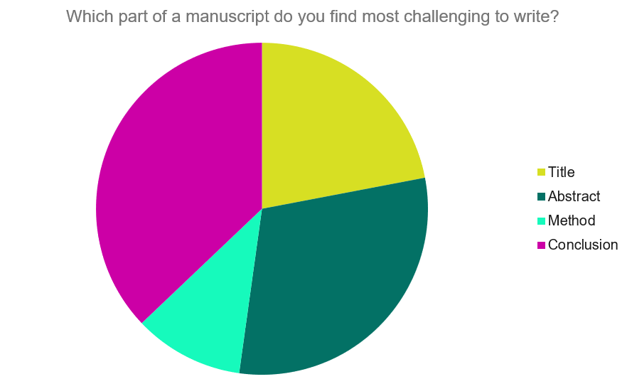
Our Early Career Researcher community tells us that the conclusion is often considered the most difficult aspect of a manuscript to write. To help, this guide provides questions to ask yourself, a basic structure to model your discussion off of and examples from published manuscripts.
Questions to ask yourself:
- Was my hypothesis correct?
- If my hypothesis is partially correct or entirely different, what can be learned from the results?
- How do the conclusions reshape or add onto the existing knowledge in the field? What does previous research say about the topic?
- Why are the results important or relevant to your audience? Do they add further evidence to a scientific consensus or disprove prior studies?
- How can future research build on these observations? What are the key experiments that must be done?
- What is the “take-home” message you want your reader to leave with?
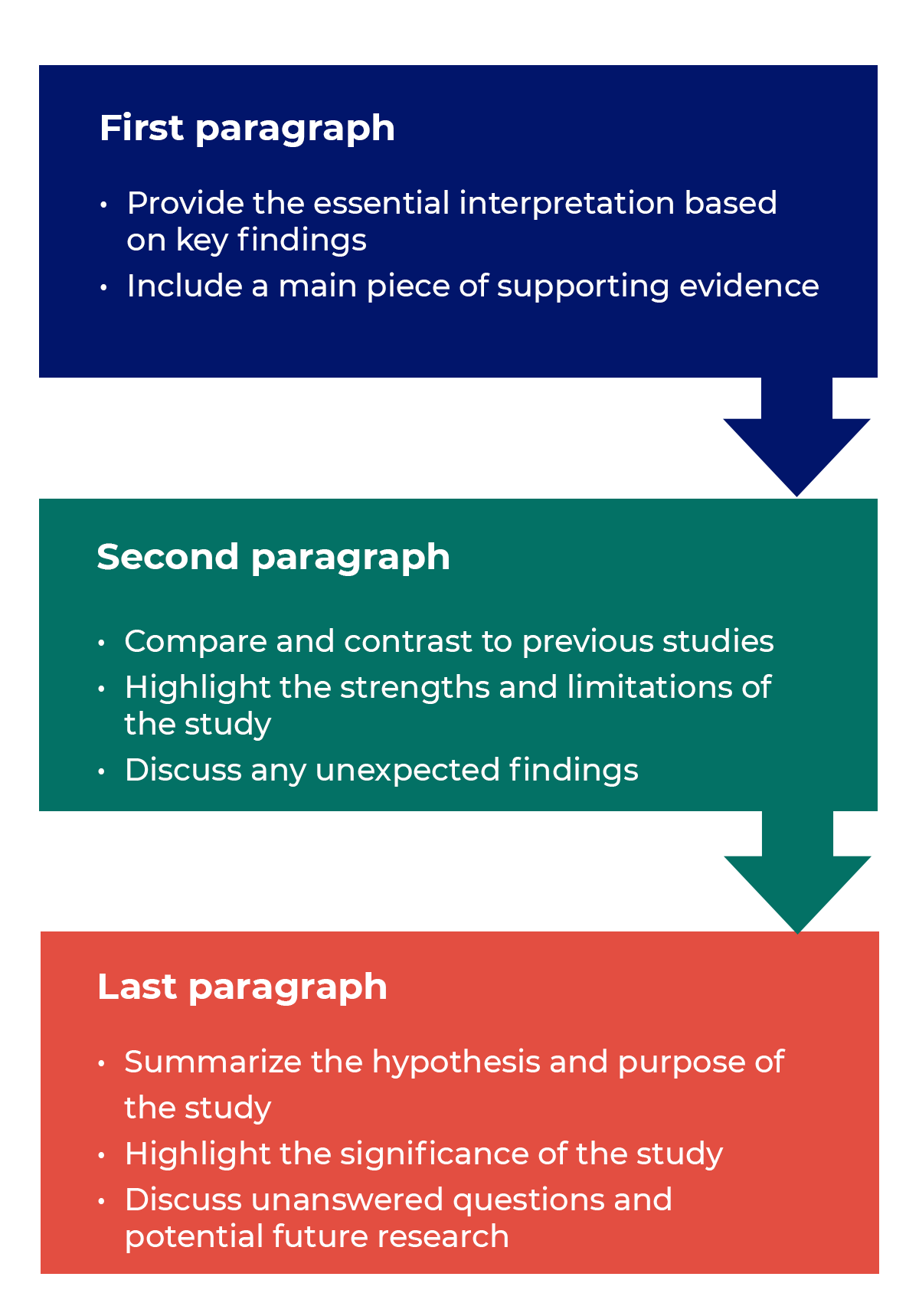
How to structure a discussion
Trying to fit a complete discussion into a single paragraph can add unnecessary stress to the writing process. If possible, you’ll want to give yourself two or three paragraphs to give the reader a comprehensive understanding of your study as a whole. Here’s one way to structure an effective discussion:
Writing Tips
While the above sections can help you brainstorm and structure your discussion, there are many common mistakes that writers revert to when having difficulties with their paper. Writing a discussion can be a delicate balance between summarizing your results, providing proper context for your research and avoiding introducing new information. Remember that your paper should be both confident and honest about the results!

- Read the journal’s guidelines on the discussion and conclusion sections. If possible, learn about the guidelines before writing the discussion to ensure you’re writing to meet their expectations.
- Begin with a clear statement of the principal findings. This will reinforce the main take-away for the reader and set up the rest of the discussion.
- Explain why the outcomes of your study are important to the reader. Discuss the implications of your findings realistically based on previous literature, highlighting both the strengths and limitations of the research.
- State whether the results prove or disprove your hypothesis. If your hypothesis was disproved, what might be the reasons?
- Introduce new or expanded ways to think about the research question. Indicate what next steps can be taken to further pursue any unresolved questions.
- If dealing with a contemporary or ongoing problem, such as climate change, discuss possible consequences if the problem is avoided.
- Be concise. Adding unnecessary detail can distract from the main findings.

Don’t
- Rewrite your abstract. Statements with “we investigated” or “we studied” generally do not belong in the discussion.
- Include new arguments or evidence not previously discussed. Necessary information and evidence should be introduced in the main body of the paper.
- Apologize. Even if your research contains significant limitations, don’t undermine your authority by including statements that doubt your methodology or execution.
- Shy away from speaking on limitations or negative results. Including limitations and negative results will give readers a complete understanding of the presented research. Potential limitations include sources of potential bias, threats to internal or external validity, barriers to implementing an intervention and other issues inherent to the study design.
- Overstate the importance of your findings. Making grand statements about how a study will fully resolve large questions can lead readers to doubt the success of the research.
Snippets of Effective Discussions:
Consumer-based actions to reduce plastic pollution in rivers: A multi-criteria decision analysis approach
Identifying reliable indicators of fitness in polar bears
- How to Write a Great Title
- How to Write an Abstract
- How to Write Your Methods
- How to Report Statistics
- How to Edit Your Work
The contents of the Peer Review Center are also available as a live, interactive training session, complete with slides, talking points, and activities. …
The contents of the Writing Center are also available as a live, interactive training session, complete with slides, talking points, and activities. …
There’s a lot to consider when deciding where to submit your work. Learn how to choose a journal that will help your study reach its audience, while reflecting your values as a researcher…
- USC Libraries
- Research Guides
Organizing Your Social Sciences Research Paper
- 8. The Discussion
- Purpose of Guide
- Design Flaws to Avoid
- Independent and Dependent Variables
- Glossary of Research Terms
- Reading Research Effectively
- Narrowing a Topic Idea
- Broadening a Topic Idea
- Extending the Timeliness of a Topic Idea
- Academic Writing Style
- Applying Critical Thinking
- Choosing a Title
- Making an Outline
- Paragraph Development
- Research Process Video Series
- Executive Summary
- The C.A.R.S. Model
- Background Information
- The Research Problem/Question
- Theoretical Framework
- Citation Tracking
- Content Alert Services
- Evaluating Sources
- Primary Sources
- Secondary Sources
- Tiertiary Sources
- Scholarly vs. Popular Publications
- Qualitative Methods
- Quantitative Methods
- Insiderness
- Using Non-Textual Elements
- Limitations of the Study
- Common Grammar Mistakes
- Writing Concisely
- Avoiding Plagiarism
- Footnotes or Endnotes?
- Further Readings
- Generative AI and Writing
- USC Libraries Tutorials and Other Guides
- Bibliography
The purpose of the discussion section is to interpret and describe the significance of your findings in relation to what was already known about the research problem being investigated and to explain any new understanding or insights that emerged as a result of your research. The discussion will always connect to the introduction by way of the research questions or hypotheses you posed and the literature you reviewed, but the discussion does not simply repeat or rearrange the first parts of your paper; the discussion clearly explains how your study advanced the reader's understanding of the research problem from where you left them at the end of your review of prior research.
Annesley, Thomas M. “The Discussion Section: Your Closing Argument.” Clinical Chemistry 56 (November 2010): 1671-1674; Peacock, Matthew. “Communicative Moves in the Discussion Section of Research Articles.” System 30 (December 2002): 479-497.
Importance of a Good Discussion
The discussion section is often considered the most important part of your research paper because it:
- Most effectively demonstrates your ability as a researcher to think critically about an issue, to develop creative solutions to problems based upon a logical synthesis of the findings, and to formulate a deeper, more profound understanding of the research problem under investigation;
- Presents the underlying meaning of your research, notes possible implications in other areas of study, and explores possible improvements that can be made in order to further develop the concerns of your research;
- Highlights the importance of your study and how it can contribute to understanding the research problem within the field of study;
- Presents how the findings from your study revealed and helped fill gaps in the literature that had not been previously exposed or adequately described; and,
- Engages the reader in thinking critically about issues based on an evidence-based interpretation of findings; it is not governed strictly by objective reporting of information.
Annesley Thomas M. “The Discussion Section: Your Closing Argument.” Clinical Chemistry 56 (November 2010): 1671-1674; Bitchener, John and Helen Basturkmen. “Perceptions of the Difficulties of Postgraduate L2 Thesis Students Writing the Discussion Section.” Journal of English for Academic Purposes 5 (January 2006): 4-18; Kretchmer, Paul. Fourteen Steps to Writing an Effective Discussion Section. San Francisco Edit, 2003-2008.
Structure and Writing Style
I. General Rules
These are the general rules you should adopt when composing your discussion of the results :
- Do not be verbose or repetitive; be concise and make your points clearly
- Avoid the use of jargon or undefined technical language
- Follow a logical stream of thought; in general, interpret and discuss the significance of your findings in the same sequence you described them in your results section [a notable exception is to begin by highlighting an unexpected result or a finding that can grab the reader's attention]
- Use the present verb tense, especially for established facts; however, refer to specific works or prior studies in the past tense
- If needed, use subheadings to help organize your discussion or to categorize your interpretations into themes
II. The Content
The content of the discussion section of your paper most often includes :
- Explanation of results : Comment on whether or not the results were expected for each set of findings; go into greater depth to explain findings that were unexpected or especially profound. If appropriate, note any unusual or unanticipated patterns or trends that emerged from your results and explain their meaning in relation to the research problem.
- References to previous research : Either compare your results with the findings from other studies or use the studies to support a claim. This can include re-visiting key sources already cited in your literature review section, or, save them to cite later in the discussion section if they are more important to compare with your results instead of being a part of the general literature review of prior research used to provide context and background information. Note that you can make this decision to highlight specific studies after you have begun writing the discussion section.
- Deduction : A claim for how the results can be applied more generally. For example, describing lessons learned, proposing recommendations that can help improve a situation, or highlighting best practices.
- Hypothesis : A more general claim or possible conclusion arising from the results [which may be proved or disproved in subsequent research]. This can be framed as new research questions that emerged as a consequence of your analysis.
III. Organization and Structure
Keep the following sequential points in mind as you organize and write the discussion section of your paper:
- Think of your discussion as an inverted pyramid. Organize the discussion from the general to the specific, linking your findings to the literature, then to theory, then to practice [if appropriate].
- Use the same key terms, narrative style, and verb tense [present] that you used when describing the research problem in your introduction.
- Begin by briefly re-stating the research problem you were investigating and answer all of the research questions underpinning the problem that you posed in the introduction.
- Describe the patterns, principles, and relationships shown by each major findings and place them in proper perspective. The sequence of this information is important; first state the answer, then the relevant results, then cite the work of others. If appropriate, refer the reader to a figure or table to help enhance the interpretation of the data [either within the text or as an appendix].
- Regardless of where it's mentioned, a good discussion section includes analysis of any unexpected findings. This part of the discussion should begin with a description of the unanticipated finding, followed by a brief interpretation as to why you believe it appeared and, if necessary, its possible significance in relation to the overall study. If more than one unexpected finding emerged during the study, describe each of them in the order they appeared as you gathered or analyzed the data. As noted, the exception to discussing findings in the same order you described them in the results section would be to begin by highlighting the implications of a particularly unexpected or significant finding that emerged from the study, followed by a discussion of the remaining findings.
- Before concluding the discussion, identify potential limitations and weaknesses if you do not plan to do so in the conclusion of the paper. Comment on their relative importance in relation to your overall interpretation of the results and, if necessary, note how they may affect the validity of your findings. Avoid using an apologetic tone; however, be honest and self-critical [e.g., in retrospect, had you included a particular question in a survey instrument, additional data could have been revealed].
- The discussion section should end with a concise summary of the principal implications of the findings regardless of their significance. Give a brief explanation about why you believe the findings and conclusions of your study are important and how they support broader knowledge or understanding of the research problem. This can be followed by any recommendations for further research. However, do not offer recommendations which could have been easily addressed within the study. This would demonstrate to the reader that you have inadequately examined and interpreted the data.
IV. Overall Objectives
The objectives of your discussion section should include the following: I. Reiterate the Research Problem/State the Major Findings
Briefly reiterate the research problem or problems you are investigating and the methods you used to investigate them, then move quickly to describe the major findings of the study. You should write a direct, declarative, and succinct proclamation of the study results, usually in one paragraph.
II. Explain the Meaning of the Findings and Why They are Important
No one has thought as long and hard about your study as you have. Systematically explain the underlying meaning of your findings and state why you believe they are significant. After reading the discussion section, you want the reader to think critically about the results and why they are important. You don’t want to force the reader to go through the paper multiple times to figure out what it all means. If applicable, begin this part of the section by repeating what you consider to be your most significant or unanticipated finding first, then systematically review each finding. Otherwise, follow the general order you reported the findings presented in the results section.
III. Relate the Findings to Similar Studies
No study in the social sciences is so novel or possesses such a restricted focus that it has absolutely no relation to previously published research. The discussion section should relate your results to those found in other studies, particularly if questions raised from prior studies served as the motivation for your research. This is important because comparing and contrasting the findings of other studies helps to support the overall importance of your results and it highlights how and in what ways your study differs from other research about the topic. Note that any significant or unanticipated finding is often because there was no prior research to indicate the finding could occur. If there is prior research to indicate this, you need to explain why it was significant or unanticipated. IV. Consider Alternative Explanations of the Findings
It is important to remember that the purpose of research in the social sciences is to discover and not to prove . When writing the discussion section, you should carefully consider all possible explanations for the study results, rather than just those that fit your hypothesis or prior assumptions and biases. This is especially important when describing the discovery of significant or unanticipated findings.
V. Acknowledge the Study’s Limitations
It is far better for you to identify and acknowledge your study’s limitations than to have them pointed out by your professor! Note any unanswered questions or issues your study could not address and describe the generalizability of your results to other situations. If a limitation is applicable to the method chosen to gather information, then describe in detail the problems you encountered and why. VI. Make Suggestions for Further Research
You may choose to conclude the discussion section by making suggestions for further research [as opposed to offering suggestions in the conclusion of your paper]. Although your study can offer important insights about the research problem, this is where you can address other questions related to the problem that remain unanswered or highlight hidden issues that were revealed as a result of conducting your research. You should frame your suggestions by linking the need for further research to the limitations of your study [e.g., in future studies, the survey instrument should include more questions that ask..."] or linking to critical issues revealed from the data that were not considered initially in your research.
NOTE: Besides the literature review section, the preponderance of references to sources is usually found in the discussion section . A few historical references may be helpful for perspective, but most of the references should be relatively recent and included to aid in the interpretation of your results, to support the significance of a finding, and/or to place a finding within a particular context. If a study that you cited does not support your findings, don't ignore it--clearly explain why your research findings differ from theirs.
V. Problems to Avoid
- Do not waste time restating your results . Should you need to remind the reader of a finding to be discussed, use "bridge sentences" that relate the result to the interpretation. An example would be: “In the case of determining available housing to single women with children in rural areas of Texas, the findings suggest that access to good schools is important...," then move on to further explaining this finding and its implications.
- As noted, recommendations for further research can be included in either the discussion or conclusion of your paper, but do not repeat your recommendations in the both sections. Think about the overall narrative flow of your paper to determine where best to locate this information. However, if your findings raise a lot of new questions or issues, consider including suggestions for further research in the discussion section.
- Do not introduce new results in the discussion section. Be wary of mistaking the reiteration of a specific finding for an interpretation because it may confuse the reader. The description of findings [results section] and the interpretation of their significance [discussion section] should be distinct parts of your paper. If you choose to combine the results section and the discussion section into a single narrative, you must be clear in how you report the information discovered and your own interpretation of each finding. This approach is not recommended if you lack experience writing college-level research papers.
- Use of the first person pronoun is generally acceptable. Using first person singular pronouns can help emphasize a point or illustrate a contrasting finding. However, keep in mind that too much use of the first person can actually distract the reader from the main points [i.e., I know you're telling me this--just tell me!].
Analyzing vs. Summarizing. Department of English Writing Guide. George Mason University; Discussion. The Structure, Format, Content, and Style of a Journal-Style Scientific Paper. Department of Biology. Bates College; Hess, Dean R. "How to Write an Effective Discussion." Respiratory Care 49 (October 2004); Kretchmer, Paul. Fourteen Steps to Writing to Writing an Effective Discussion Section. San Francisco Edit, 2003-2008; The Lab Report. University College Writing Centre. University of Toronto; Sauaia, A. et al. "The Anatomy of an Article: The Discussion Section: "How Does the Article I Read Today Change What I Will Recommend to my Patients Tomorrow?” The Journal of Trauma and Acute Care Surgery 74 (June 2013): 1599-1602; Research Limitations & Future Research . Lund Research Ltd., 2012; Summary: Using it Wisely. The Writing Center. University of North Carolina; Schafer, Mickey S. Writing the Discussion. Writing in Psychology course syllabus. University of Florida; Yellin, Linda L. A Sociology Writer's Guide . Boston, MA: Allyn and Bacon, 2009.
Writing Tip
Don’t Over-Interpret the Results!
Interpretation is a subjective exercise. As such, you should always approach the selection and interpretation of your findings introspectively and to think critically about the possibility of judgmental biases unintentionally entering into discussions about the significance of your work. With this in mind, be careful that you do not read more into the findings than can be supported by the evidence you have gathered. Remember that the data are the data: nothing more, nothing less.
MacCoun, Robert J. "Biases in the Interpretation and Use of Research Results." Annual Review of Psychology 49 (February 1998): 259-287; Ward, Paulet al, editors. The Oxford Handbook of Expertise . Oxford, UK: Oxford University Press, 2018.
Another Writing Tip
Don't Write Two Results Sections!
One of the most common mistakes that you can make when discussing the results of your study is to present a superficial interpretation of the findings that more or less re-states the results section of your paper. Obviously, you must refer to your results when discussing them, but focus on the interpretation of those results and their significance in relation to the research problem, not the data itself.
Azar, Beth. "Discussing Your Findings." American Psychological Association gradPSYCH Magazine (January 2006).
Yet Another Writing Tip
Avoid Unwarranted Speculation!
The discussion section should remain focused on the findings of your study. For example, if the purpose of your research was to measure the impact of foreign aid on increasing access to education among disadvantaged children in Bangladesh, it would not be appropriate to speculate about how your findings might apply to populations in other countries without drawing from existing studies to support your claim or if analysis of other countries was not a part of your original research design. If you feel compelled to speculate, do so in the form of describing possible implications or explaining possible impacts. Be certain that you clearly identify your comments as speculation or as a suggestion for where further research is needed. Sometimes your professor will encourage you to expand your discussion of the results in this way, while others don’t care what your opinion is beyond your effort to interpret the data in relation to the research problem.
- << Previous: Using Non-Textual Elements
- Next: Limitations of the Study >>
- Last Updated: Jun 18, 2024 10:45 AM
- URL: https://libguides.usc.edu/writingguide
How to Write the Discussion Section of a Research Paper
The discussion section of a research paper analyzes and interprets the findings, provides context, compares them with previous studies, identifies limitations, and suggests future research directions.
Updated on September 15, 2023

Structure your discussion section right, and you’ll be cited more often while doing a greater service to the scientific community. So, what actually goes into the discussion section? And how do you write it?
The discussion section of your research paper is where you let the reader know how your study is positioned in the literature, what to take away from your paper, and how your work helps them. It can also include your conclusions and suggestions for future studies.
First, we’ll define all the parts of your discussion paper, and then look into how to write a strong, effective discussion section for your paper or manuscript.
Discussion section: what is it, what it does
The discussion section comes later in your paper, following the introduction, methods, and results. The discussion sets up your study’s conclusions. Its main goals are to present, interpret, and provide a context for your results.
What is it?
The discussion section provides an analysis and interpretation of the findings, compares them with previous studies, identifies limitations, and suggests future directions for research.
This section combines information from the preceding parts of your paper into a coherent story. By this point, the reader already knows why you did your study (introduction), how you did it (methods), and what happened (results). In the discussion, you’ll help the reader connect the ideas from these sections.
Why is it necessary?
The discussion provides context and interpretations for the results. It also answers the questions posed in the introduction. While the results section describes your findings, the discussion explains what they say. This is also where you can describe the impact or implications of your research.
Adds context for your results
Most research studies aim to answer a question, replicate a finding, or address limitations in the literature. These goals are first described in the introduction. However, in the discussion section, the author can refer back to them to explain how the study's objective was achieved.
Shows what your results actually mean and real-world implications
The discussion can also describe the effect of your findings on research or practice. How are your results significant for readers, other researchers, or policymakers?
What to include in your discussion (in the correct order)
A complete and effective discussion section should at least touch on the points described below.
Summary of key findings
The discussion should begin with a brief factual summary of the results. Concisely overview the main results you obtained.
Begin with key findings with supporting evidence
Your results section described a list of findings, but what message do they send when you look at them all together?
Your findings were detailed in the results section, so there’s no need to repeat them here, but do provide at least a few highlights. This will help refresh the reader’s memory and help them focus on the big picture.
Read the first paragraph of the discussion section in this article (PDF) for an example of how to start this part of your paper. Notice how the authors break down their results and follow each description sentence with an explanation of why each finding is relevant.
State clearly and concisely
Following a clear and direct writing style is especially important in the discussion section. After all, this is where you will make some of the most impactful points in your paper. While the results section often contains technical vocabulary, such as statistical terms, the discussion section lets you describe your findings more clearly.
Interpretation of results
Once you’ve given your reader an overview of your results, you need to interpret those results. In other words, what do your results mean? Discuss the findings’ implications and significance in relation to your research question or hypothesis.
Analyze and interpret your findings
Look into your findings and explore what’s behind them or what may have caused them. If your introduction cited theories or studies that could explain your findings, use these sources as a basis to discuss your results.
For example, look at the second paragraph in the discussion section of this article on waggling honey bees. Here, the authors explore their results based on information from the literature.
Unexpected or contradictory results
Sometimes, your findings are not what you expect. Here’s where you describe this and try to find a reason for it. Could it be because of the method you used? Does it have something to do with the variables analyzed? Comparing your methods with those of other similar studies can help with this task.
Context and comparison with previous work
Refer to related studies to place your research in a larger context and the literature. Compare and contrast your findings with existing literature, highlighting similarities, differences, and/or contradictions.
How your work compares or contrasts with previous work
Studies with similar findings to yours can be cited to show the strength of your findings. Information from these studies can also be used to help explain your results. Differences between your findings and others in the literature can also be discussed here.
How to divide this section into subsections
If you have more than one objective in your study or many key findings, you can dedicate a separate section to each of these. Here’s an example of this approach. You can see that the discussion section is divided into topics and even has a separate heading for each of them.
Limitations
Many journals require you to include the limitations of your study in the discussion. Even if they don’t, there are good reasons to mention these in your paper.
Why limitations don’t have a negative connotation
A study’s limitations are points to be improved upon in future research. While some of these may be flaws in your method, many may be due to factors you couldn’t predict.
Examples include time constraints or small sample sizes. Pointing this out will help future researchers avoid or address these issues. This part of the discussion can also include any attempts you have made to reduce the impact of these limitations, as in this study .
How limitations add to a researcher's credibility
Pointing out the limitations of your study demonstrates transparency. It also shows that you know your methods well and can conduct a critical assessment of them.
Implications and significance
The final paragraph of the discussion section should contain the take-home messages for your study. It can also cite the “strong points” of your study, to contrast with the limitations section.
Restate your hypothesis
Remind the reader what your hypothesis was before you conducted the study.
How was it proven or disproven?
Identify your main findings and describe how they relate to your hypothesis.
How your results contribute to the literature
Were you able to answer your research question? Or address a gap in the literature?
Future implications of your research
Describe the impact that your results may have on the topic of study. Your results may show, for instance, that there are still limitations in the literature for future studies to address. There may be a need for studies that extend your findings in a specific way. You also may need additional research to corroborate your findings.
Sample discussion section
This fictitious example covers all the aspects discussed above. Your actual discussion section will probably be much longer, but you can read this to get an idea of everything your discussion should cover.
Our results showed that the presence of cats in a household is associated with higher levels of perceived happiness by its human occupants. These findings support our hypothesis and demonstrate the association between pet ownership and well-being.
The present findings align with those of Bao and Schreer (2016) and Hardie et al. (2023), who observed greater life satisfaction in pet owners relative to non-owners. Although the present study did not directly evaluate life satisfaction, this factor may explain the association between happiness and cat ownership observed in our sample.
Our findings must be interpreted in light of some limitations, such as the focus on cat ownership only rather than pets as a whole. This may limit the generalizability of our results.
Nevertheless, this study had several strengths. These include its strict exclusion criteria and use of a standardized assessment instrument to investigate the relationships between pets and owners. These attributes bolster the accuracy of our results and reduce the influence of confounding factors, increasing the strength of our conclusions. Future studies may examine the factors that mediate the association between pet ownership and happiness to better comprehend this phenomenon.
This brief discussion begins with a quick summary of the results and hypothesis. The next paragraph cites previous research and compares its findings to those of this study. Information from previous studies is also used to help interpret the findings. After discussing the results of the study, some limitations are pointed out. The paper also explains why these limitations may influence the interpretation of results. Then, final conclusions are drawn based on the study, and directions for future research are suggested.
How to make your discussion flow naturally
If you find writing in scientific English challenging, the discussion and conclusions are often the hardest parts of the paper to write. That’s because you’re not just listing up studies, methods, and outcomes. You’re actually expressing your thoughts and interpretations in words.
- How formal should it be?
- What words should you use, or not use?
- How do you meet strict word limits, or make it longer and more informative?
Always give it your best, but sometimes a helping hand can, well, help. Getting a professional edit can help clarify your work’s importance while improving the English used to explain it. When readers know the value of your work, they’ll cite it. We’ll assign your study to an expert editor knowledgeable in your area of research. Their work will clarify your discussion, helping it to tell your story. Find out more about AJE Editing.

Adam Goulston, PsyD, MS, MBA, MISD, ELS
Science Marketing Consultant
See our "Privacy Policy"
Ensure your structure and ideas are consistent and clearly communicated
Pair your Premium Editing with our add-on service Presubmission Review for an overall assessment of your manuscript.

An official website of the United States government
The .gov means it’s official. Federal government websites often end in .gov or .mil. Before sharing sensitive information, make sure you’re on a federal government site.
The site is secure. The https:// ensures that you are connecting to the official website and that any information you provide is encrypted and transmitted securely.
- Publications
- Account settings
Preview improvements coming to the PMC website in October 2024. Learn More or Try it out now .
- Advanced Search
- Journal List
- BMC Med Res Methodol
Writing a discussion section: how to integrate substantive and statistical expertise
Michael höfler.
1 Institute of Clinical Psychology and Psychotherapy, Technische Universität Dresden, Dresden, Germany
5 Chair of Clinical Psychology and Behavioural Neuroscience, Institute of Clinical Psychology and Psychotherapy, Technische Universität Dresden, Dresden, Germany
2 Behavioral Epidemiology, Institute of Clinical Psychology and Psychotherapy, Technische Universität Dresden, Dresden, Germany
Sebastian Trautmann
Robert miller.
3 Faculty of Psychology, Technische Universität Dresden, Dresden, Germany
4 Department of Medical Epidemiology and Biostatistics, Karolinska Institute, Stockholm, Sweden
Associated Data
Not applicable.
When discussing results medical research articles often tear substantive and statistical (methodical) contributions apart, just as if both were independent. Consequently, reasoning on bias tends to be vague, unclear and superficial. This can lead to over-generalized, too narrow and misleading conclusions, especially for causal research questions.
To get the best possible conclusion, substantive and statistical expertise have to be integrated on the basis of reasonable assumptions. While statistics should raise questions on the mechanisms that have presumably created the data, substantive knowledge should answer them. Building on the related principle of Bayesian thinking, we make seven specific and four general proposals on writing a discussion section.
Misinterpretation could be reduced if authors explicitly discussed what can be concluded under which assumptions. Informed on the resulting conditional conclusions other researchers may, according to their knowledge and beliefs, follow a particular conclusion or, based on other conditions, arrive at another one. This could foster both an improved debate and a better understanding of the mechanisms behind the data and should therefore enable researchers to better address bias in future studies.
After a research article has presented the substantive background, the methods and the results, the discussion section assesses the validity of results and draws conclusions by interpreting them. The discussion puts the results into a broader context and reflects their implications for theoretical (e.g. etiological) and practical (e.g. interventional) purposes. As such, the discussion contains an article’s last words the reader is left with.
Common recommendations for the discussion section include general proposals for writing [ 1 ] and structuring (e.g. with a paragraph on a study’s strengths and weaknesses) [ 2 ], to avoid common statistical pitfalls (like misinterpreting non-significant findings as true null results) [ 3 ] and to “go beyond the data” when interpreting results [ 4 ]. Note that the latter includes much more than comparing an article’s results with the literature. If results and literature are consistent, this might be due to shared bias only. If they are not consistent, the question arises why inconsistency occurs – maybe because of bias acting differently across studies [ 5 – 7 ]. Recommendations like the CONSORT checklist do well in demanding all quantitative information on design, participation, compliance etc. to be reported in the methods and results section and “addressing sources of potential bias”, “limitations” and “considering other relevant evidence” in the discussion [ 8 , 9 ]. Similarly, the STROBE checklist for epidemiological research demands “a cautious overall interpretation of results” and "discussing the generalizability (external validity)" [ 10 , 11 ]. However, these guidelines do not clarify how to deal with the complex bias issue, and how to get to and report conclusions.
Consequently, suggestions on writing a discussion often remain vague by hardly addressing the role of the assumptions that have (often implicitly) been made when designing a study, analyzing the data and interpreting the results. Such assumptions involve mechanisms that have created the data and are related to sampling, measurement and treatment assignment (in observational studies common causes of factor and outcome) and, as a consequence, the bias this may produce [ 5 , 6 ]. They determine whether a result allows only an associational or a causal conclusion. Causal conclusions, if true, are of much higher relevance for etiology, prevention and intervention. However, they require much stronger assumptions. These have to be fully explicit and, therewith, essential part of the debate since they always involve subjectivity. Subjectivity is unavoidable because the mechanisms behind the data can never be fully estimated from the data themselves [ 12 ].
In this article, we argue that the conjunction of substantive and statistical (methodical) knowledge in the verbal integration of results and beliefs on mechanisms can be greatly improved in (medical) research papers. We illustrate this through the personal roles that a statistician (i.e. methods expert) and a substantive researcher should take. Doing so, we neither claim that usually just two people write a discussion, nor that one person lacks the knowledge of the other, nor that there were truly no researchers that have both kinds of expertise. As a metaphor, the division of these two roles into two persons describes the necessary integration of knowledge via the mode of a dialogue. Verbally, it addresses the finding of increased specialization of different study contributors in biomedical research. This has teared apart the two processes of statistical compilation of results and their verbal integration [ 13 ]. When this happens a statistician alone is limited to a study’s conditions (sampled population, experimental settings etc.), because he or she is unaware of the conditions’ generalizability. On the other hand, a A substantive expert alone is prone to over-generalize because he or she is not aware of the (mathematical) prerequisites for an interpretation.
The article addresses both (medical) researchers educated in basic statistics and research methods and statisticians who cooperate with them. Throughout the paper we exemplify our arguments with the finding of an association in a cross-tabulation between a binary X (factor) and a binary Y (outcome): those who are exposed to or treated with X have a statistically significantly elevated risk for Y as compared to the non-exposed or not (or otherwise) treated (for instance via the chi-squared independence test or logistic regression). Findings like this are frequent and raise the question which more profound conclusion is valid under what assumptions. Until some decades ago, statistics has largely avoided the related topic of causality and instead limited itself on describing observed distributions (here a two-by-two table between D = depression and LC = lung cancer) with well-fitting models.
We illustrate our arguments with the concrete example of the association found between the factor depression (D) and the outcome lung cancer (LC) [ 14 ]. Yet very different mechanisms could have produced such an association [ 7 ], and assumptions on these lead to the following fundamentally different conclusions (Fig. (Fig.1 1 ):
- D causes LC (e.g. because smoking might constitute “self-medication” of depression symptoms)
- LC causes D (e.g. because LC patients are demoralized by their diagnosis)
- D and LC cause each other (e.g. because the arguments in both a. and b. apply)
- D and LC are the causal consequence of the same factor(s) (e.g. poor health behaviors - HB)
- D and LC only share measurement error (e.g. because a fraction of individuals that has either depression or lung cancer denies both in self-report measures).

Different conclusions about an association between D and LC. a D causes LC, b LC causes B, c D and LC cause each other, d D and LC are associated because of a shared factor (HB), e D and LC are associated because they have correlated errors
Note that we use the example purely for illustrative purposes. We do not make substantive claims on what of a. through e. is true but show how one should reflect on mechanisms in order to find the right answer. Besides, we do not consider research on the D-LC relation apart from the finding of association [ 14 ].
Assessing which of a. through e. truly applies requires substantive assumptions on mechanisms: the temporal order of D and LC (a causal effect requires that the cause occurs before the effect), shared factors, selection processes and measurement error. Questions on related mechanisms have to be brought up by statistical consideration, while substantive reasoning has to address them. Together this yields provisional assumptions for inferring that are subject to readers’ substantive consideration and refinement. In general, the integration of prior beliefs (anything beyond the data a conclusion depends on) and the results from the data themselves is formalized by Bayesian statistics [ 15 , 16 ]. This is beyond the scope of this article, still we argue that Bayesian thinking should govern the process of drawing conclusions.
Building on this idea, we provide seven specific and four general recommendations for the cooperative process of writing a discussion. The recommendations are intended to be suggestions rather than rules. They should be subject to further refinement and adjustment to specific requirements in different fields of medical and other research. Note that the order of the points is not meant to structure a discussion’s writing (besides 1.).
Recommendations for writing a discussion section
Specific recommendations.
Consider the example on the association between D and LC. Rather than starting with an in-depth (causal) interpretation a finding should firstly be taken as what it allows inferring without doubt: Under the usual assumptions that a statistical model makes (e.g. random sampling, independence or certain correlation structure between observations [ 17 ]), the association indicates that D (strictly speaking: measuring D) predicts an elevated LC risk (strictly speaking: measuring LC) in the population that one has managed to sample (source population). Assume that the sample has been randomly drawn from primary care settings. In this case the association is useful to recommend medical doctors to better look at an individual’s LC risk in case of D. If the association has been adjusted for age and gender (conveniently through a regression model), the conclusion modifies to: If the doctor knows a patient’s age and gender (what should always be the case) D has additional value in predicting an elevated LC risk.
In the above example, a substantive researcher might want to conclude that D and LC are associated in a general population instead of just inferring to patients in primary care settings (a.). Another researcher might even take the finding as evidence for D being a causal factor in the etiology of LC, meaning that prevention of D could reduce the incidence rate of LC (in whatever target population) (b.). In both cases, the substantive researcher should insist on assessing the desired interpretation that goes beyond the data [ 4 ], but the statistician immediately needs to bring up the next point.
The explanation of all the assumptions that lead from a data result to a conclusion enables a reader to assess whether he or she agrees with the authors’ inference or not. These conditions, however, often remain incomplete or unclear, in which case the reader can hardly assess whether he or she follows a path of argumentation and, thus, shares the conclusion this path leads to.
Consider conclusion a. and suppose that, instead of representative sampling in a general population (e.g. all U.S. citizens aged 18 or above), the investigators were only able to sample in primary care settings. Extrapolating the results to another population than the source population requires what is called “external validity”, “transportability” or the absence of “selection bias” [ 18 , 19 ]. No such bias occurs if the parameter of interest is equal in the source and the target population. Note that this is a weaker condition than the common belief that the sample must represent the target population in everything . If the parameter of interest is the difference in risk for LC between cases and non-cases of D, the condition translates into: the risk difference must be equal in target and source population.
For the causal conclusion b., however, sufficient assumptions are very strict. In an RCT, the conclusion is valid under random sampling from the target population, random allocation of X, perfect compliance in X, complete participation and no measurement error in outcome (for details see [ 20 ]). In practice, on the other hand, the derivations from such conditions might sometimes be modest what may produce little bias only. For instance, non-compliance in a specific drug intake (treatment) might occur only in a few individuals to little extent through a random process (e.g. sickness of a nurse being responsible for drug dispense) and yield just small (downward) bias [ 5 ]. The conclusion of downward bias might also be justified if non-compliance does not cause anything that has a larger effect on a Y than the drug itself. Another researcher, however, could believe that non-compliance leads to taking a more effective, alternative treatment. He or she could infer upward bias instead if well-informed on the line of argument.
In practice, researchers frequently use causal language yet without mentioning any assumptions. This does not imply that they truly have a causal effect in mind, often causal and associational wordings are carelessly used in synonymous way. For example, concluding “depression increases the risk of lung cancer” constitutes already causal wording because it implies that a change in the depression status would change the cancer risk. Associational language like “lung cancer risk is elevated if depression occurs”, however, would allow for an elevated lung cancer risk in depression cases just because LC and D share some causes (“inducing” or “removing” depression would not change the cancer risk here).
Often, it is unclear where the path of argumentation from assumptions to a conclusion leads when alternative assumptions are made. Consider again bias due to selection. A different effect in target and source population occurs if effect-modifying variables distribute differently in both populations. Accordingly, the statistician should ask which variables influence the effect of interest, and whether these can be assumed to distribute equally in the source population and the target population. The substantive researcher might answer that the causal risk difference between D and LC likely increases with age. Given that this is true, and if elder individuals have been oversampled (e.g. because elderly are over-represented in primary care settings), both together would conclude that sampling has led to over-estimation (despite other factors, Fig. Fig.2 2 ).

If higher age is related to a larger effect (risk difference) of D on LC, a larger effect estimate is expected in an elder sample
However, the statistician might add, if effect modification is weak, or the difference in the age distributions is modest (e.g. mean 54 vs. 52 years), selection is unlikely to have produced large (here: upward) bias. In turn, another substantive researcher, who reads the resulting discussion, might instead assume a decrease of effect with increasing age and thus infer downward bias.
In practice, researchers should be extremely sensitive for bias due to selection if a sample has been drawn conditionally on a common consequence of factor and outcome or a variable associated with such a consequence [19 and references therein]. For instance, hospitalization might be influenced by both D and LC, and thus sampling from hospitals might introduce a false association or change an association’s sign; particularly D and LC may appear to be negatively associated although the association is positive in the general population (Fig. (Fig.3 3 ).

If hospitalization (H) is a common cause of D and LC, sampling conditionally on H can introduce a spurious association between D and LC ("conditioning on a collider")
Usually, only some kinds of bias are discussed, while the consequences of others are ignored [ 5 ]. Besides selection the main sources of bias are often measurement and confounding. If one is only interested in association, confounding is irrelevant. For causal conclusions, however, assumptions on all three kinds of bias are necessary.
Measurement error means that the measurement of a factor and/or outcome deviates from the true value, at least in some individuals. Bias due to measurement is known under many other terms that describe the reasons why such error occurs (e.g. “recall bias” and “reporting bias”). In contrast to conventional wisdom, measurement error does not always bias association and effect estimates downwards [ 5 , 6 ]. It does, for instance, if only the factor (e.g. depression) is measured with error and the errors occur independently from the outcome (e.g. lung cancer), or vice versa (“non-differential misclassification”) [22 and references therein]. However, many lung cancer cases might falsely report depression symptoms (e.g. to express need for care). Such false positives (non-cases of depression classified as cases) may also occur in non-cases of lung cancer but to a lesser extent (a special case of “differential misclassification”). Here, bias might be upward as well. Importantly, false positives cause larger bias than false negatives (non-cases of depression falsely classified as depression cases) as long as the relative frequency of a factor is lower than 50% [ 21 ]. Therefore, they should receive more attention in discussion. If measurement error occurs in depression and lung cancer, the direction of bias also depends on the correlation between both errors [ 21 ].
Note that what is in line with common standards of “good” measurement (e.g. a Kappa value measuring validity or reliability of 0.7) might anyway produce large bias. This applies to estimates of prevalence, association and effect. The reason is that while indices of measurement are one-dimensional, bias depends on two parameters (sensitivity and specificity) [ 21 , 22 ]. Moreover, estimates of such indices are often extrapolated to different kinds of populations (typically from a clinical to general population), what may be inadequate. Note that the different kinds of bias often interact, e.g. bias due to measurement might depend on selection (e.g. measurement error might differ between a clinical and a general population) [ 5 , 6 ].
Assessment of bias due to confounding variables (roughly speaking: common causes of factor and outcome) requires assumptions on the entire system of variables that affect both factor and outcome. For example, D and LC might share several causes such as stressful life events or socioeconomic status. If these influence D and LC with the same effect direction, this leads to overestimation, otherwise (different effect directions) the causal effect is underestimated. In the medical field, many unfavorable conditions may be positively related. If this holds true for all common factors of D and LC, upward bias can be assumed. However, not all confounders have to be taken into account. Within the framework of “causal graphs”, the “backdoor criterion” [ 7 ] provides a graphical rule for sets of confounders to be sufficient when adjusted for. Practically, such a causal graph must include all factors that directly or indirectly affect both D and LC. Then, adjustment for a set of confounders that meets the “backdoor criterion” in the graph completely removes bias due to confounding. In the example of Fig. Fig.4 4 it is sufficient to adjust for Z 1 and Z 2 because this “blocks” all paths that otherwise lead backwards from D to LC. Note that fully eliminating bias due to confounding also requires that the confounders have been collected without measurement error [ 5 , 6 , 23 ]. Therefore, the advice is always to concede at least some “residual” bias and reflect on the direction this might have (could be downward if such error is not stronger related to D and LC than a confounder itself).

Causal graph for the effect of D on LC and confounders Z 1 , Z 2 and Z 3
Whereas the statistician should pinpoint to the mathematical insight of the backdoor criterion, its application requires profound substantive input and literature review. Of course, there are numerous relevant factors in the medical field. Hence, one should practically focus on those with the highest prevalence (a very seldom factor can hardly cause large bias) and large assumed effects on both X and Y.
If knowledge on any of the three kinds of bias is poor or very uncertain, researchers should admit that this adds uncertainty in a conclusion: systematic error on top of random error. In the Bayesian framework, quantitative bias analysis formalizes this through the result of larger variance in an estimate. Technically, this additional variance is introduced via the variances of distributions assigned to “bias parameters”; for instance a misclassification probability (e.g. classifying a true depression case as non-case) or the prevalence of a binary confounder and its effects on X and Y. Of course, bias analysis also changes point estimates (hopefully reducing bias considerably). Note that conventional frequentist analysis, as regarded from the Bayesian perspective, assumes that all bias parameters were zero with a probability of one [ 5 , 6 , 23 ]. The only exceptions (bias addressed in conventional analyses) are adjustment on variables to hopefully reduce bias due to confounding and weighting the individuals (according to variables related to participation) to take into account bias due to selection.
If the substantive investigator understands the processes of selection, measurement and confounding only poorly, such strict analysis numerically reveals that little to nothing is known on the effect of X on Y, no matter how large an observed association and a sample (providing small random error) may be [ 5 , 6 , 23 ]). This insight has to be brought up by the statistician. Although such an analysis is complicated, itself very sensitive to how it is conducted [ 5 , 6 ] and rarely done, the Bayesian thinking behind it forces researchers to better understand the processes behind the data. Otherwise, he or she cannot make any assumptions and, in turn, no conclusion on causality.
Usually articles end with statements that only go little further than the always true but never informative statement “more research is needed”. Moreover, larger samples and better measurements are frequently proposed. If an association has been found, a RCT or other interventional study is usually proposed to investigate causality. In our example, this recommendation disregards that: (1) onset of D might have a different effect on LC risk than an intervention against D (the effect of onset cannot be investigated in any interventional study), (2) the effects of onset and intervention concern different populations (those without vs. those with depression), (3) an intervention effect depends on the mode of intervention [ 24 ], and (4) (applying the backdoor criterion) a well-designed observational study may approximatively yield the same result as a randomized study would [ 25 – 27 ]. If the effect of “removing” depression is actually of interest, one could propose an RCT that investigates the effect of treating depression in a strictly defined way and in a strictly defined population (desirably in all who meet the criteria of depression). Ideally, this population is sampled randomly, and non-participants and dropouts are investigated with respect to assumed effect-modifiers (differences in their distributions between participants and non-participants can then be addressed e.g. by weighting [ 27 ]). In a non-randomized study, one should collect variables supposed to meet the backdoor-criterion with the best instruments possible.
General recommendations
Yet when considering 1) through 7); i.e. carefully reflecting on the mechanisms that have created the data, discussions on statistical results can be very misleading, because the basic statistical methods are mis-interpreted or inadequately worded.
A common pitfall is to consider the lack of evidence for the alternative hypothesis (e.g. association between D and LC) as evidence for the null hypothesis (no association). In fact, such inference requires an a-priori calculated sample-size to ensure that the type-two error probability does not exceed a pre-specified limit (typically 20% or 10%, given the other necessary assumptions, e.g. on the true magnitude of association). Otherwise, the type-two error is unknown and in practice often large. This may put a “false negative result” into the scientific public that turns out to be “unreplicable” – what would be falsely interpreted as part of the “replication crisis”. Such results are neither positive nor negative but uninformative . In this case, the wording “there is no evidence for an association” is adequate because it does not claim that there is no association.
Frequently, it remains unclear which hypotheses have been a-priori specified and which have been brought up only after some data analysis. This, of course, is scientific malpractice because it does not enable the readership to assess the random error emerging from explorative data analysis. Accordingly, the variance of results across statistical methods is often misused to filter out the analysis that yields a significant result (“ p -hacking”, [ 28 ]). Pre-planned tests (via writing a grant) leave at least less room for p-hacking because they specify a-priori which analysis is to be conducted.
On the other hand, post-hoc analyses can be extremely useful for identifying unexpected phenomena and creating new hypotheses. Verbalization in the discussion section should therefore sharply separate between conclusions from hypothesis testing and new hypotheses created from data exploration. The distinction is profound, since a newly proposed hypothesis just makes a new claim. Suggesting new hypotheses cannot be wrong, this can only be inefficient if many hypotheses turn out to be wrong. Therefore, we suggest proposing only a limited number of new hypotheses that appear promising to stimulate further research and scientific progress. They are to be confirmed or falsified with future studies. A present discussion, however, should yet explicate the testable predictions a new hypothesis entails, and how a future study should be designed to keep bias in related analyses as small as possible.
Confidence intervals address the problem of reducing results to the dichotomy of significant and non-significant through providing a range of values that are compatible with the data at the given confidence level, usually 95% [ 29 ].
This is also addressed by Bayesian statistics that allows calculating what frequentist p -values are often misinterpreted to be: the probability that the alternative (or null) hypothesis is true [ 17 ]. Moreover, one can calculate how likely it is that the parameter lies within any specified range (e.g. the risk difference being greater than .05, a lower boundary for practical significance) [ 15 , 16 ]. To gain these benefits, one needs to specify how the parameter of interest (e.g. causal risk difference between D and LC) is distributed before inspecting the data. In Bayesian statistics (unlike frequentist statistics) a parameter is a random number that expresses prior beliefs via a “prior distribution”. Such a “prior” is combined with the data result to a “posterior distribution”. This integrates both sources of information.
Note that confidence intervals also can be interpreted from the Bayesian perspective (then called “credibility interval”). This assumes that all parameter values were equally likely (uniformly distributed, strictly speaking) before analyzing the data [ 5 , 6 , 20 ].
Testing just for a non-zero association can only yield evidence for an association deviating from zero. A better indicator for the true impact of an effect/association for clinical, economic, political, or research purposes is its magnitude. If an association between D and LC after adjusting for age and gender has been discovered, then the knowledge of D has additional value in predicting an elevated LC probability beyond age and gender. However, there may be many other factors that stronger predict LC and thus should receive higher priority in a doctor’s assessment. Besides, if an association is small, it may yet be explained by modest (upward) bias. Especially large samples often yield significant results with little practical value. The p -value does not measure strength of association [ 17 ]. For instance, in a large sample, a Pearson correlation between two dimensional variables could equal 0.1 only but with a p -value <.001. A further problem arises if the significance threshold of .05 is weakened post-hoc to allow for “statistical trends” ( p between .05 and .10) because a result has “failed to reach significance” (this wording claims that there is truly an association. If this was known, no research would be necessary).
It is usually the statistician’s job to insist not only on removing the attention from pure statistical significance to confidence intervals or even Bayesian interpretation, but also to point out the necessity of a meaningful cutoff for practical significance. The substantive researcher then has to provide this cutoff.
Researchers should not draw conclusions that have not been explicitly tested for. For example, one may have found a positive association between D and LC (e.g. p = .049), but this association is not significant (e.g. p = .051), when adjusting for “health behavior”. This does not imply that “health behavior” “explains” the association (yet fully). The difference in magnitude of association in both analyses compared here (without and with adjustment on HB) may be very small and the difference in p -values (“borderline significance” after adjustment) likely to emerge from random error. This often applies to larger differences in p as well.
Investigators, however, might find patterns in their results that they consider worth mentioning for creating hypotheses. In the example above, adding the words “in the sample”, would clarify that they refer just to the difference of two point estimates . By default, “association” in hypotheses testing should mean “statistically significant association” (explorative analyses should instead refer to “suggestive associations”).
Conclusions
Some issues of discussing results not mentioned yet appear to require only substantive reasoning. For instance, Bradford Hill’s consideration on “plausibility” claims that a causal effect is more likely, if it is in line with biological (substantive) knowledge, or if a dose-response relation has been found [ 30 ]. However, the application of these considerations itself depends on the trueness of assumptions. For instance, bias might act differently across the dose of exposure (e.g. larger measurement error in outcome among those with higher dosage). As a consequence, a pattern observed across dose may mask a true or pretend a wrong dose-response relation [ 30 ]. This again has to be brought up by statistical expertise.
There are, however, some practical issues that hinder the cooperation we suggest. First, substantive researchers often feel discomfort when urged to make assumptions on the mechanisms behind the data, presumably because they fear to be wrong. Here, the statistician needs to insist: “If you are unable to make any assumptions, you cannot conclude anything!” And: “As a scientist you have to understand the processes that create your data.” See [ 31 ] for practical advice on how to arrive at meaningful assumptions.
Second, statisticians have long been skeptical against causal inference. Still, most of them focus solely on describing observed data with distributional models, probably because estimating causal effects has long been regarded as unfeasible with scientific methods. Training in causality remains rather new, since strict mathematical methods have been developed only in the last decades [ 7 ].
The cooperation could be improved if education in both fields focused on the insight that one cannot succeed without the other. Academic education should demonstrate that in-depth conclusions from data unavoidably involve prior beliefs. Such education should say: Data do not “speak for themselves”, because they “speak” only ambiguously and little, since they have been filtered through various biases [ 32 ]. The subjectivity introduced by addressing bias, however, unsettles many researchers. On the other hand, conventional frequentist statistics just pretends to be objective. Instead of accepting the variety of possible assumptions, it makes the absurd assumption of “no bias with probability of one”. Or it avoids causal conclusions at all if no randomized study is possible. This limits science to investigating just associations for all factors that can never be randomized (e.g. onset of depression). However, the alternative of Bayesian statistics and thinking are themselves prone to fundamental cognitive biases which should as well be subject of interdisciplinary teaching [ 33 ].
Readers may take this article as an invitation to read further papers’ discussions differently while evaluating our claims. Rather than sharing a provided conclusion (or not) they could ask themselves whether a discussion enables them to clearly specify why they share it (or not). If the result is uncertainty, this might motivate them to write their next discussion differently. The proposals made in this article could help shifting scientific debates to where they belong. Rather than arguing on misunderstandings caused by ambiguity in a conclusion’s assumptions one should argue on the assumptions themselves.
Acknowledgements
We acknowledge support by the German Research Foundation and the Open Access Publication Funds of the TU Dresden. We wish to thank Pia Grabbe and Helen Steiner for language editing and the cited authors for their outstanding work that our proposals build on.
John Venz is funded by the German Federal Ministry of Education and Research (BMBF) project no. 01ER1303 and 01ER1703. He has contributed to this manuscript outside of time funded by these projects.
Availability of data and materials
Abbreviations.
| D | depression |
| HB | health behavior |
| LC | lung cancer |
| RCT | randomized clinical trial |
| X | factor variable |
| Y | outcome variable |
Authors’ contributions
MH and RM had the initial idea on the article. MH has taken the lead in writing. JV has contributed to the statistical parts, especially the Bayesian aspects. RM has refined the paragraphs on statistical inference. ST joined later and has added many clarifications related to the perspective of the substantive researcher. All authors have contributed to the final wording of all sections and the article’s revision. All authors read and approved the final manuscript.
Ethics approval and consent to participate
Consent for publication, competing interests.
The authors declare that they have no competing interests.
Publisher’s Note
Springer Nature remains neutral with regard to jurisdictional claims in published maps and institutional affiliations.
How Do I Write the Discussion Chapter?
Reflecting on and Comparing Your Data, Recognising the Strengths and Limitations
- First Online: 19 October 2023
Cite this chapter

- Sue Reeves ORCID: orcid.org/0000-0002-3017-0559 3 &
- Bartek Buczkowski ORCID: orcid.org/0000-0002-4146-3664 4
481 Accesses
The Discussion chapter brings an opportunity to write an academic argument that contains a detailed critical evaluation and analysis of your research findings. This chapter addresses the purpose and critical nature of the discussion, contains a guide to selecting key results to discuss, and details how best to structure the discussion with subsections and paragraphs. We also present a list of points to do and avoid when writing the discussion together with a Discussion chapter checklist.
This is a preview of subscription content, log in via an institution to check access.
Access this chapter
- Available as EPUB and PDF
- Read on any device
- Instant download
- Own it forever
- Compact, lightweight edition
- Dispatched in 3 to 5 business days
- Free shipping worldwide - see info
Tax calculation will be finalised at checkout
Purchases are for personal use only
Institutional subscriptions
Braun V, Clarke V (2013) Successful qualitative research: a practical guide for beginners. SAGE Publications, London
Google Scholar
McGregor SLT (2018) Understanding and evaluating research: a critical guide. SAGE Publications, Los Angeles, CA
Book Google Scholar
PLOS (2023) Author resources. How to write discussions and conclusions. Accessed Mar 3, 2023, from https://plos.org/resource/how-to-write-conclusions/ . Accessed 3 Mar 2023
Further Reading
Cottrell S (2017) Critical thinking skills: effective analysis, argument and reflection, 3rd edn. Palgrave, London
Download references
Author information
Authors and affiliations.
University of Roehampton, London, UK
Manchester Metropolitan University, Manchester, UK
Bartek Buczkowski
You can also search for this author in PubMed Google Scholar
Rights and permissions
Reprints and permissions
Copyright information
© 2023 The Author(s), under exclusive license to Springer Nature Switzerland AG
About this chapter
Reeves, S., Buczkowski, B. (2023). How Do I Write the Discussion Chapter?. In: Mastering Your Dissertation. Springer, Cham. https://doi.org/10.1007/978-3-031-41911-9_9
Download citation
DOI : https://doi.org/10.1007/978-3-031-41911-9_9
Published : 19 October 2023
Publisher Name : Springer, Cham
Print ISBN : 978-3-031-41910-2
Online ISBN : 978-3-031-41911-9
eBook Packages : Biomedical and Life Sciences Biomedical and Life Sciences (R0)
Share this chapter
Anyone you share the following link with will be able to read this content:
Sorry, a shareable link is not currently available for this article.
Provided by the Springer Nature SharedIt content-sharing initiative
- Publish with us
Policies and ethics
- Find a journal
- Track your research

- Langson Library
- Science Library
- Grunigen Medical Library
- Law Library
- Connect From Off-Campus
- Accessibility
- Gateway Study Center
Email this link
Writing a scientific paper.
- Writing a lab report
- INTRODUCTION
Writing a "good" discussion section
"discussion and conclusions checklist" from: how to write a good scientific paper. chris a. mack. spie. 2018., peer review.
- LITERATURE CITED
- Bibliography of guides to scientific writing and presenting
- Presentations
- Lab Report Writing Guides on the Web
This is is usually the hardest section to write. You are trying to bring out the true meaning of your data without being too long. Do not use words to conceal your facts or reasoning. Also do not repeat your results, this is a discussion.
- Present principles, relationships and generalizations shown by the results
- Point out exceptions or lack of correlations. Define why you think this is so.
- Show how your results agree or disagree with previously published works
- Discuss the theoretical implications of your work as well as practical applications
- State your conclusions clearly. Summarize your evidence for each conclusion.
- Discuss the significance of the results
- Evidence does not explain itself; the results must be presented and then explained.
- Typical stages in the discussion: summarizing the results, discussing whether results are expected or unexpected, comparing these results to previous work, interpreting and explaining the results (often by comparison to a theory or model), and hypothesizing about their generality.
- Discuss any problems or shortcomings encountered during the course of the work.
- Discuss possible alternate explanations for the results.
- Avoid: presenting results that are never discussed; presenting discussion that does not relate to any of the results; presenting results and discussion in chronological order rather than logical order; ignoring results that do not support the conclusions; drawing conclusions from results without logical arguments to back them up.
CONCLUSIONS
- Provide a very brief summary of the Results and Discussion.
- Emphasize the implications of the findings, explaining how the work is significant and providing the key message(s) the author wishes to convey.
- Provide the most general claims that can be supported by the evidence.
- Provide a future perspective on the work.
- Avoid: repeating the abstract; repeating background information from the Introduction; introducing new evidence or new arguments not found in the Results and Discussion; repeating the arguments made in the Results and Discussion; failing to address all of the research questions set out in the Introduction.
WHAT HAPPENS AFTER I COMPLETE MY PAPER?
The peer review process is the quality control step in the publication of ideas. Papers that are submitted to a journal for publication are sent out to several scientists (peers) who look carefully at the paper to see if it is "good science". These reviewers then recommend to the editor of a journal whether or not a paper should be published. Most journals have publication guidelines. Ask for them and follow them exactly. Peer reviewers examine the soundness of the materials and methods section. Are the materials and methods used written clearly enough for another scientist to reproduce the experiment? Other areas they look at are: originality of research, significance of research question studied, soundness of the discussion and interpretation, correct spelling and use of technical terms, and length of the article.
- << Previous: RESULTS
- Next: LITERATURE CITED >>
- Last Updated: Aug 4, 2023 9:33 AM
- URL: https://guides.lib.uci.edu/scientificwriting
Off-campus? Please use the Software VPN and choose the group UCIFull to access licensed content. For more information, please Click here
Software VPN is not available for guests, so they may not have access to some content when connecting from off-campus.
- Affiliate Program

- UNITED STATES
- 台灣 (TAIWAN)
- TÜRKIYE (TURKEY)
- Academic Editing Services
- - Research Paper
- - Journal Manuscript
- - Dissertation
- - College & University Assignments
- Admissions Editing Services
- - Application Essay
- - Personal Statement
- - Recommendation Letter
- - Cover Letter
- - CV/Resume
- Business Editing Services
- - Business Documents
- - Report & Brochure
- - Website & Blog
- Writer Editing Services
- - Script & Screenplay
- Our Editors
- Client Reviews
- Editing & Proofreading Prices
- Wordvice Points
- Partner Discount
- Plagiarism Checker
- APA Citation Generator
- MLA Citation Generator
- Chicago Citation Generator
- Vancouver Citation Generator
- - APA Style
- - MLA Style
- - Chicago Style
- - Vancouver Style
- Writing & Editing Guide
- Academic Resources
- Admissions Resources
How to Write a Discussion Section for a Research Paper
We’ve talked about several useful writing tips that authors should consider while drafting or editing their research papers. In particular, we’ve focused on figures and legends , as well as the Introduction , Methods , and Results . Now that we’ve addressed the more technical portions of your journal manuscript, let’s turn to the analytical segments of your research article. In this article, we’ll provide tips on how to write a strong Discussion section that best portrays the significance of your research contributions.
What is the Discussion section of a research paper?
In a nutshell, your Discussion fulfills the promise you made to readers in your Introduction . At the beginning of your paper, you tell us why we should care about your research. You then guide us through a series of intricate images and graphs that capture all the relevant data you collected during your research. We may be dazzled and impressed at first, but none of that matters if you deliver an anti-climactic conclusion in the Discussion section!
Are you feeling pressured? Don’t worry. To be honest, you will edit the Discussion section of your manuscript numerous times. After all, in as little as one to two paragraphs ( Nature ‘s suggestion based on their 3,000-word main body text limit), you have to explain how your research moves us from point A (issues you raise in the Introduction) to point B (our new understanding of these matters). You must also recommend how we might get to point C (i.e., identify what you think is the next direction for research in this field). That’s a lot to say in two paragraphs!
So, how do you do that? Let’s take a closer look.
What should I include in the Discussion section?
As we stated above, the goal of your Discussion section is to answer the questions you raise in your Introduction by using the results you collected during your research . The content you include in the Discussions segment should include the following information:
- Remind us why we should be interested in this research project.
- Describe the nature of the knowledge gap you were trying to fill using the results of your study.
- Don’t repeat your Introduction. Instead, focus on why this particular study was needed to fill the gap you noticed and why that gap needed filling in the first place.
- Mainly, you want to remind us of how your research will increase our knowledge base and inspire others to conduct further research.
- Clearly tell us what that piece of missing knowledge was.
- Answer each of the questions you asked in your Introduction and explain how your results support those conclusions.
- Make sure to factor in all results relevant to the questions (even if those results were not statistically significant).
- Focus on the significance of the most noteworthy results.
- If conflicting inferences can be drawn from your results, evaluate the merits of all of them.
- Don’t rehash what you said earlier in the Results section. Rather, discuss your findings in the context of answering your hypothesis. Instead of making statements like “[The first result] was this…,” say, “[The first result] suggests [conclusion].”
- Do your conclusions line up with existing literature?
- Discuss whether your findings agree with current knowledge and expectations.
- Keep in mind good persuasive argument skills, such as explaining the strengths of your arguments and highlighting the weaknesses of contrary opinions.
- If you discovered something unexpected, offer reasons. If your conclusions aren’t aligned with current literature, explain.
- Address any limitations of your study and how relevant they are to interpreting your results and validating your findings.
- Make sure to acknowledge any weaknesses in your conclusions and suggest room for further research concerning that aspect of your analysis.
- Make sure your suggestions aren’t ones that should have been conducted during your research! Doing so might raise questions about your initial research design and protocols.
- Similarly, maintain a critical but unapologetic tone. You want to instill confidence in your readers that you have thoroughly examined your results and have objectively assessed them in a way that would benefit the scientific community’s desire to expand our knowledge base.
- Recommend next steps.
- Your suggestions should inspire other researchers to conduct follow-up studies to build upon the knowledge you have shared with them.
- Keep the list short (no more than two).
How to Write the Discussion Section
The above list of what to include in the Discussion section gives an overall idea of what you need to focus on throughout the section. Below are some tips and general suggestions about the technical aspects of writing and organization that you might find useful as you draft or revise the contents we’ve outlined above.
Technical writing elements
- Embrace active voice because it eliminates the awkward phrasing and wordiness that accompanies passive voice.
- Use the present tense, which should also be employed in the Introduction.
- Sprinkle with first person pronouns if needed, but generally, avoid it. We want to focus on your findings.
- Maintain an objective and analytical tone.
Discussion section organization
- Keep the same flow across the Results, Methods, and Discussion sections.
- We develop a rhythm as we read and parallel structures facilitate our comprehension. When you organize information the same way in each of these related parts of your journal manuscript, we can quickly see how a certain result was interpreted and quickly verify the particular methods used to produce that result.
- Notice how using parallel structure will eliminate extra narration in the Discussion part since we can anticipate the flow of your ideas based on what we read in the Results segment. Reducing wordiness is important when you only have a few paragraphs to devote to the Discussion section!
- Within each subpart of a Discussion, the information should flow as follows: (A) conclusion first, (B) relevant results and how they relate to that conclusion and (C) relevant literature.
- End with a concise summary explaining the big-picture impact of your study on our understanding of the subject matter. At the beginning of your Discussion section, you stated why this particular study was needed to fill the gap you noticed and why that gap needed filling in the first place. Now, it is time to end with “how your research filled that gap.”
Discussion Part 1: Summarizing Key Findings
Begin the Discussion section by restating your statement of the problem and briefly summarizing the major results. Do not simply repeat your findings. Rather, try to create a concise statement of the main results that directly answer the central research question that you stated in the Introduction section . This content should not be longer than one paragraph in length.
Many researchers struggle with understanding the precise differences between a Discussion section and a Results section . The most important thing to remember here is that your Discussion section should subjectively evaluate the findings presented in the Results section, and in relatively the same order. Keep these sections distinct by making sure that you do not repeat the findings without providing an interpretation.
Phrase examples: Summarizing the results
- The findings indicate that …
- These results suggest a correlation between A and B …
- The data present here suggest that …
- An interpretation of the findings reveals a connection between…
Discussion Part 2: Interpreting the Findings
What do the results mean? It may seem obvious to you, but simply looking at the figures in the Results section will not necessarily convey to readers the importance of the findings in answering your research questions.
The exact structure of interpretations depends on the type of research being conducted. Here are some common approaches to interpreting data:
- Identifying correlations and relationships in the findings
- Explaining whether the results confirm or undermine your research hypothesis
- Giving the findings context within the history of similar research studies
- Discussing unexpected results and analyzing their significance to your study or general research
- Offering alternative explanations and arguing for your position
Organize the Discussion section around key arguments, themes, hypotheses, or research questions or problems. Again, make sure to follow the same order as you did in the Results section.
Discussion Part 3: Discussing the Implications
In addition to providing your own interpretations, show how your results fit into the wider scholarly literature you surveyed in the literature review section. This section is called the implications of the study . Show where and how these results fit into existing knowledge, what additional insights they contribute, and any possible consequences that might arise from this knowledge, both in the specific research topic and in the wider scientific domain.
Questions to ask yourself when dealing with potential implications:
- Do your findings fall in line with existing theories, or do they challenge these theories or findings? What new information do they contribute to the literature, if any? How exactly do these findings impact or conflict with existing theories or models?
- What are the practical implications on actual subjects or demographics?
- What are the methodological implications for similar studies conducted either in the past or future?
Your purpose in giving the implications is to spell out exactly what your study has contributed and why researchers and other readers should be interested.
Phrase examples: Discussing the implications of the research
- These results confirm the existing evidence in X studies…
- The results are not in line with the foregoing theory that…
- This experiment provides new insights into the connection between…
- These findings present a more nuanced understanding of…
- While previous studies have focused on X, these results demonstrate that Y.
Step 4: Acknowledging the limitations
All research has study limitations of one sort or another. Acknowledging limitations in methodology or approach helps strengthen your credibility as a researcher. Study limitations are not simply a list of mistakes made in the study. Rather, limitations help provide a more detailed picture of what can or cannot be concluded from your findings. In essence, they help temper and qualify the study implications you listed previously.
Study limitations can relate to research design, specific methodological or material choices, or unexpected issues that emerged while you conducted the research. Mention only those limitations directly relate to your research questions, and explain what impact these limitations had on how your study was conducted and the validity of any interpretations.
Possible types of study limitations:
- Insufficient sample size for statistical measurements
- Lack of previous research studies on the topic
- Methods/instruments/techniques used to collect the data
- Limited access to data
- Time constraints in properly preparing and executing the study
After discussing the study limitations, you can also stress that your results are still valid. Give some specific reasons why the limitations do not necessarily handicap your study or narrow its scope.
Phrase examples: Limitations sentence beginners
- “There may be some possible limitations in this study.”
- “The findings of this study have to be seen in light of some limitations.”
- “The first limitation is the…The second limitation concerns the…”
- “The empirical results reported herein should be considered in the light of some limitations.”
- “This research, however, is subject to several limitations.”
- “The primary limitation to the generalization of these results is…”
- “Nonetheless, these results must be interpreted with caution and a number of limitations should be borne in mind.”
Discussion Part 5: Giving Recommendations for Further Research
Based on your interpretation and discussion of the findings, your recommendations can include practical changes to the study or specific further research to be conducted to clarify the research questions. Recommendations are often listed in a separate Conclusion section , but often this is just the final paragraph of the Discussion section.
Suggestions for further research often stem directly from the limitations outlined. Rather than simply stating that “further research should be conducted,” provide concrete specifics for how future can help answer questions that your research could not.
Phrase examples: Recommendation sentence beginners
- Further research is needed to establish …
- There is abundant space for further progress in analyzing…
- A further study with more focus on X should be done to investigate…
- Further studies of X that account for these variables must be undertaken.
Consider Receiving Professional Language Editing
As you edit or draft your research manuscript, we hope that you implement these guidelines to produce a more effective Discussion section. And after completing your draft, don’t forget to submit your work to a professional proofreading and English editing service like Wordvice, including our manuscript editing service for paper editing , cover letter editing , SOP editing , and personal statement proofreading services. Language editors not only proofread and correct errors in grammar, punctuation, mechanics, and formatting but also improve terms and revise phrases so they read more naturally. Wordvice is an industry leader in providing high-quality revision for all types of academic documents.
For additional information about how to write a strong research paper, make sure to check out our full research writing series !
Wordvice Writing Resources
- How to Write a Research Paper Introduction
- Which Verb Tenses to Use in a Research Paper
- How to Write an Abstract for a Research Paper
- How to Write a Research Paper Title
- Useful Phrases for Academic Writing
- Common Transition Terms in Academic Papers
- Active and Passive Voice in Research Papers
- 100+ Verbs That Will Make Your Research Writing Amazing
- Tips for Paraphrasing in Research Papers
Additional Academic Resources
- Guide for Authors. (Elsevier)
- How to Write the Results Section of a Research Paper. (Bates College)
- Structure of a Research Paper. (University of Minnesota Biomedical Library)
- How to Choose a Target Journal (Springer)
- How to Write Figures and Tables (UNC Writing Center)
How to Start a Discussion Section in Research? [with Examples]
The examples below are from 72,017 full-text PubMed research papers that I analyzed in order to explore common ways to start writing the Discussion section.
Research papers included in this analysis were selected at random from those uploaded to PubMed Central between the years 2016 and 2021. Note that I used the BioC API to download the data (see the References section below).
Examples of how to start writing the Discussion section
In the Discussion section, you should explain the meaning of your results, their importance, and implications. [for more information, see: How to Write & Publish a Research Paper: Step-by-Step Guide ]
The Discussion section can:
1. Start by restating the study objective
“ The purpose of this study was to investigate the relationship between muscle synergies and motion primitives of the upper limb motions.” Taken from the Discussion section of this article on PubMed
“ The main objective of this study was to identify trajectories of autonomy.” Taken from the Discussion section of this article on PubMed
“ In the present study, we investigated the whole brain regional homogeneity in patients with melancholic MDD and non-melancholic MDD at rest . “ Taken from the Discussion section of this article on PubMed
2. Start by mentioning the main finding
“ We found that autocracy and democracy have acted as peaks in an evolutionary landscape of possible modes of institutional arrangements.” Taken from the Discussion section of this article on PubMed
“ In this study, we demonstrated that the neural mechanisms of rhythmic movements and skilled movements are similar.” Taken from the Discussion section of this article on PubMed
“ The results of this study show that older adults are a diverse group concerning their activities on the Internet.” Taken from the Discussion section of this article on PubMed
3. Start by pointing out the strength of the study
“ To our knowledge, this investigation is by far the largest epidemiological study employing real-time PCR to study periodontal pathogens in subgingival plaque.” Taken from the Discussion section of this article on PubMed
“ This is the first human subject research using the endoscopic hemoglobin oxygen saturation imaging technology for patients with aero-digestive tract cancers or adenomas.” Taken from the Discussion section of this article on PubMed
“ In this work, we introduced a new real-time flow imaging method and systematically demonstrated its effectiveness with both flow phantom experiments and in vivo experiments.” Taken from the Discussion section of this article on PubMed
Most used words at the start of the Discussion
Here are the top 10 phrases used to start a discussion section in our dataset:
| Rank | Phrase | Percent of occurrences |
|---|---|---|
| 1 | “In this study,…” | 4.48% |
| 2 | “In the present study,…” | 1.66% |
| 3 | “To our knowledge,…” | 0.73% |
| 4 | “To the best of our knowledge,…” | 0.51% |
| 5 | “In the current study,…” | 0.38% |
| 6 | “The aim of this study was…” | 0.38% |
| 7 | “This is the first study to…” | 0.28% |
| 8 | “The purpose of this study was to…” | 0.22% |
| 9 | “The results of the present study…” | 0.14% |
| 10 | “The aim of the present study was…” | 0.11% |
- Comeau DC, Wei CH, Islamaj Doğan R, and Lu Z. PMC text mining subset in BioC: about 3 million full text articles and growing, Bioinformatics , btz070, 2019.
Further reading
- How Long Should the Discussion Section Be? Data from 61,517 Examples
- How to Write & Publish a Research Paper: Step-by-Step Guide
- “I” & “We” in Academic Writing: Examples from 9,830 Studies
- Walden University
- Faculty Portal
General Research Paper Guidelines: Discussion
Discussion section.
The overall purpose of a research paper’s discussion section is to evaluate and interpret results, while explaining both the implications and limitations of your findings. Per APA (2020) guidelines, this section requires you to “examine, interpret, and qualify the results and draw inferences and conclusions from them” (p. 89). Discussion sections also require you to detail any new insights, think through areas for future research, highlight the work that still needs to be done to further your topic, and provide a clear conclusion to your research paper. In a good discussion section, you should do the following:
- Clearly connect the discussion of your results to your introduction, including your central argument, thesis, or problem statement.
- Provide readers with a critical thinking through of your results, answering the “so what?” question about each of your findings. In other words, why is this finding important?
- Detail how your research findings might address critical gaps or problems in your field
- Compare your results to similar studies’ findings
- Provide the possibility of alternative interpretations, as your goal as a researcher is to “discover” and “examine” and not to “prove” or “disprove.” Instead of trying to fit your results into your hypothesis, critically engage with alternative interpretations to your results.
For more specific details on your Discussion section, be sure to review Sections 3.8 (pp. 89-90) and 3.16 (pp. 103-104) of your 7 th edition APA manual
*Box content adapted from:
University of Southern California (n.d.). Organizing your social sciences research paper: 8 the discussion . https://libguides.usc.edu/writingguide/discussion
Limitations
Limitations of generalizability or utility of findings, often over which the researcher has no control, should be detailed in your Discussion section. Including limitations for your reader allows you to demonstrate you have thought critically about your given topic, understood relevant literature addressing your topic, and chosen the methodology most appropriate for your research. It also allows you an opportunity to suggest avenues for future research on your topic. An effective limitations section will include the following:
- Detail (a) sources of potential bias, (b) possible imprecision of measures, (c) other limitations or weaknesses of the study, including any methodological or researcher limitations.
- Sample size: In quantitative research, if a sample size is too small, it is more difficult to generalize results.
- Lack of available/reliable data : In some cases, data might not be available or reliable, which will ultimately affect the overall scope of your research. Use this as an opportunity to explain areas for future study.
- Lack of prior research on your study topic: In some cases, you might find that there is very little or no similar research on your study topic, which hinders the credibility and scope of your own research. If this is the case, use this limitation as an opportunity to call for future research. However, make sure you have done a thorough search of the available literature before making this claim.
- Flaws in measurement of data: Hindsight is 20/20, and you might realize after you have completed your research that the data tool you used actually limited the scope or results of your study in some way. Again, acknowledge the weakness and use it as an opportunity to highlight areas for future study.
- Limits of self-reported data: In your research, you are assuming that any participants will be honest and forthcoming with responses or information they provide to you. Simply acknowledging this assumption as a possible limitation is important in your research.
- Access: Most research requires that you have access to people, documents, organizations, etc.. However, for various reasons, access is sometimes limited or denied altogether. If this is the case, you will want to acknowledge access as a limitation to your research.
- Time: Choosing a research focus that is narrow enough in scope to finish in a given time period is important. If such limitations of time prevent you from certain forms of research, access, or study designs, acknowledging this time restraint is important. Acknowledging such limitations is important, as they can point other researchers to areas that require future study.
- Potential Bias: All researchers have some biases, so when reading and revising your draft, pay special attention to the possibilities for bias in your own work. Such bias could be in the form you organized people, places, participants, or events. They might also exist in the method you selected or the interpretation of your results. Acknowledging such bias is an important part of the research process.
- Language Fluency: On occasion, researchers or research participants might have language fluency issues, which could potentially hinder results or how effectively you interpret results. If this is an issue in your research, make sure to acknowledge it in your limitations section.
University of Southern California (n.d.). Organizing your social sciences research paper: Limitations of the study . https://libguides.usc.edu/writingguide/limitations
In many research papers, the conclusion, like the limitations section, is folded into the larger discussion section. If you are unsure whether to include the conclusion as part of your discussion or as a separate section, be sure to defer to the assignment instructions or ask your instructor.
The conclusion is important, as it is specifically designed to highlight your research’s larger importance outside of the specific results of your study. Your conclusion section allows you to reiterate the main findings of your study, highlight their importance, and point out areas for future research. Based on the scope of your paper, your conclusion could be anywhere from one to three paragraphs long. An effective conclusion section should include the following:
- Describe the possibilities for continued research on your topic, including what might be improved, adapted, or added to ensure useful and informed future research.
- Provide a detailed account of the importance of your findings
- Reiterate why your problem is important, detail how your interpretation of results impacts the subfield of study, and what larger issues both within and outside of your field might be affected from such results
University of Southern California (n.d.). Organizing your social sciences research paper: 9. the conclusion . https://libguides.usc.edu/writingguide/conclusion
- Previous Page: Results
- Next Page: References
- Office of Student Disability Services
Walden Resources
Departments.
- Academic Residencies
- Academic Skills
- Career Planning and Development
- Customer Care Team
- Field Experience
- Military Services
- Student Success Advising
- Writing Skills
Centers and Offices
- Center for Social Change
- Office of Academic Support and Instructional Services
- Office of Degree Acceleration
- Office of Research and Doctoral Services
- Office of Student Affairs
Student Resources
- Doctoral Writing Assessment
- Form & Style Review
- Quick Answers
- ScholarWorks
- SKIL Courses and Workshops
- Walden Bookstore
- Walden Catalog & Student Handbook
- Student Safety/Title IX
- Legal & Consumer Information
- Website Terms and Conditions
- Cookie Policy
- Accessibility
- Accreditation
- State Authorization
- Net Price Calculator
- Contact Walden
Walden University is a member of Adtalem Global Education, Inc. www.adtalem.com Walden University is certified to operate by SCHEV © 2024 Walden University LLC. All rights reserved.
Organizing Academic Research Papers: 8. The Discussion
- Purpose of Guide
- Design Flaws to Avoid
- Glossary of Research Terms
- Narrowing a Topic Idea
- Broadening a Topic Idea
- Extending the Timeliness of a Topic Idea
- Academic Writing Style
- Choosing a Title
- Making an Outline
- Paragraph Development
- Executive Summary
- Background Information
- The Research Problem/Question
- Theoretical Framework
- Citation Tracking
- Content Alert Services
- Evaluating Sources
- Primary Sources
- Secondary Sources
- Tertiary Sources
- What Is Scholarly vs. Popular?
- Qualitative Methods
- Quantitative Methods
- Using Non-Textual Elements
- Limitations of the Study
- Common Grammar Mistakes
- Avoiding Plagiarism
- Footnotes or Endnotes?
- Further Readings
- Annotated Bibliography
- Dealing with Nervousness
- Using Visual Aids
- Grading Someone Else's Paper
- How to Manage Group Projects
- Multiple Book Review Essay
- Reviewing Collected Essays
- About Informed Consent
- Writing Field Notes
- Writing a Policy Memo
- Writing a Research Proposal
- Acknowledgements
The purpose of the discussion is to interpret and describe the significance of your findings in light of what was already known about the research problem being investigated, and to explain any new understanding or fresh insights about the problem after you've taken the findings into consideration. The discussion will always connect to the introduction by way of the research questions or hypotheses you posed and the literature you reviewed, but it does not simply repeat or rearrange the introduction; the discussion should always explain how your study has moved the reader's understanding of the research problem forward from where you left them at the end of the introduction.
Importance of a Good Discussion
This section is often considered the most important part of a research paper because it most effectively demonstrates your ability as a researcher to think critically about an issue, to develop creative solutions to problems based on the findings, and to formulate a deeper, more profound understanding of the research problem you are studying.
The discussion section is where you explore the underlying meaning of your research , its possible implications in other areas of study, and the possible improvements that can be made in order to further develop the concerns of your research.
This is the section where you need to present the importance of your study and how it may be able to contribute to and/or fill existing gaps in the field. If appropriate, the discussion section is also where you state how the findings from your study revealed new gaps in the literature that had not been previously exposed or adequately described.
This part of the paper is not strictly governed by objective reporting of information but, rather, it is where you can engage in creative thinking about issues through evidence-based interpretation of findings. This is where you infuse your results with meaning.
Kretchmer, Paul. Fourteen Steps to Writing to Writing an Effective Discussion Section . San Francisco Edit, 2003-2008.
Structure and Writing Style
I. General Rules
These are the general rules you should adopt when composing your discussion of the results :
- Do not be verbose or repetitive.
- Be concise and make your points clearly.
- Avoid using jargon.
- Follow a logical stream of thought.
- Use the present verb tense, especially for established facts; however, refer to specific works and references in the past tense.
- If needed, use subheadings to help organize your presentation or to group your interpretations into themes.
II. The Content
The content of the discussion section of your paper most often includes :
- Explanation of results : comment on whether or not the results were expected and present explanations for the results; go into greater depth when explaining findings that were unexpected or especially profound. If appropriate, note any unusual or unanticipated patterns or trends that emerged from your results and explain their meaning.
- References to previous research : compare your results with the findings from other studies, or use the studies to support a claim. This can include re-visiting key sources already cited in your literature review section, or, save them to cite later in the discussion section if they are more important to compare with your results than being part of the general research you cited to provide context and background information.
- Deduction : a claim for how the results can be applied more generally. For example, describing lessons learned, proposing recommendations that can help improve a situation, or recommending best practices.
- Hypothesis : a more general claim or possible conclusion arising from the results [which may be proved or disproved in subsequent research].
III. Organization and Structure
Keep the following sequential points in mind as you organize and write the discussion section of your paper:
- Think of your discussion as an inverted pyramid. Organize the discussion from the general to the specific, linking your findings to the literature, then to theory, then to practice [if appropriate].
- Use the same key terms, mode of narration, and verb tense [present] that you used when when describing the research problem in the introduction.
- Begin by briefly re-stating the research problem you were investigating and answer all of the research questions underpinning the problem that you posed in the introduction.
- Describe the patterns, principles, and relationships shown by each major findings and place them in proper perspective. The sequencing of providing this information is important; first state the answer, then the relevant results, then cite the work of others. If appropriate, refer the reader to a figure or table to help enhance the interpretation of the data. The order of interpreting each major finding should be in the same order as they were described in your results section.
- A good discussion section includes analysis of any unexpected findings. This paragraph should begin with a description of the unexpected finding, followed by a brief interpretation as to why you believe it appeared and, if necessary, its possible significance in relation to the overall study. If more than one unexpected finding emerged during the study, describe each them in the order they appeared as you gathered the data.
- Before concluding the discussion, identify potential limitations and weaknesses. Comment on their relative importance in relation to your overall interpretation of the results and, if necessary, note how they may affect the validity of the findings. Avoid using an apologetic tone; however, be honest and self-critical.
- The discussion section should end with a concise summary of the principal implications of the findings regardless of statistical significance. Give a brief explanation about why you believe the findings and conclusions of your study are important and how they support broader knowledge or understanding of the research problem. This can be followed by any recommendations for further research. However, do not offer recommendations which could have been easily addressed within the study. This demonstrates to the reader you have inadequately examined and interpreted the data.
IV. Overall Objectives
The objectives of your discussion section should include the following: I. Reiterate the Research Problem/State the Major Findings
Briefly reiterate for your readers the research problem or problems you are investigating and the methods you used to investigate them, then move quickly to describe the major findings of the study. You should write a direct, declarative, and succinct proclamation of the study results.
II. Explain the Meaning of the Findings and Why They are Important
No one has thought as long and hard about your study as you have. Systematically explain the meaning of the findings and why you believe they are important. After reading the discussion section, you want the reader to think about the results [“why hadn’t I thought of that?”]. You don’t want to force the reader to go through the paper multiple times to figure out what it all means. Begin this part of the section by repeating what you consider to be your most important finding first.
III. Relate the Findings to Similar Studies
No study is so novel or possesses such a restricted focus that it has absolutely no relation to other previously published research. The discussion section should relate your study findings to those of other studies, particularly if questions raised by previous studies served as the motivation for your study, the findings of other studies support your findings [which strengthens the importance of your study results], and/or they point out how your study differs from other similar studies. IV. Consider Alternative Explanations of the Findings
It is important to remember that the purpose of research is to discover and not to prove . When writing the discussion section, you should carefully consider all possible explanations for the study results, rather than just those that fit your prior assumptions or biases.
V. Acknowledge the Study’s Limitations
It is far better for you to identify and acknowledge your study’s limitations than to have them pointed out by your professor! Describe the generalizability of your results to other situations, if applicable to the method chosen, then describe in detail problems you encountered in the method(s) you used to gather information. Note any unanswered questions or issues your study did not address, and.... VI. Make Suggestions for Further Research
Although your study may offer important insights about the research problem, other questions related to the problem likely remain unanswered. Moreover, some unanswered questions may have become more focused because of your study. You should make suggestions for further research in the discussion section.
NOTE: Besides the literature review section, the preponderance of references to sources in your research paper are usually found in the discussion section . A few historical references may be helpful for perspective but most of the references should be relatively recent and included to aid in the interpretation of your results and/or linked to similar studies. If a study that you cited disagrees with your findings, don't ignore it--clearly explain why the study's findings differ from yours.
V. Problems to Avoid
- Do not waste entire sentences restating your results . Should you need to remind the reader of the finding to be discussed, use "bridge sentences" that relate the result to the interpretation. An example would be: “The lack of available housing to single women with children in rural areas of Texas suggests that...[then move to the interpretation of this finding].”
- Recommendations for further research can be included in either the discussion or conclusion of your paper but do not repeat your recommendations in the both sections.
- Do not introduce new results in the discussion. Be wary of mistaking the reiteration of a specific finding for an interpretation.
- Use of the first person is acceptable, but too much use of the first person may actually distract the reader from the main points.
Analyzing vs. Summarizing. Department of English Writing Guide. George Mason University; Discussion . The Structure, Format, Content, and Style of a Journal-Style Scientific Paper. Department of Biology. Bates College; Hess, Dean R. How to Write an Effective Discussion. Respiratory Care 49 (October 2004); Kretchmer, Paul. Fourteen Steps to Writing to Writing an Effective Discussion Section . San Francisco Edit, 2003-2008; The Lab Report . University College Writing Centre. University of Toronto; Summary: Using it Wisely . The Writing Center. University of North Carolina; Schafer, Mickey S. Writing the Discussion . Writing in Psychology course syllabus. University of Florida; Yellin, Linda L. A Sociology Writer's Guide. Boston, MA: Allyn and Bacon, 2009.
Writing Tip
Don’t Overinterpret the Results!
Interpretation is a subjective exercise. Therefore, be careful that you do not read more into the findings than can be supported by the evidence you've gathered. Remember that the data are the data: nothing more, nothing less.
Another Writing Tip
Don't Write Two Results Sections!
One of the most common mistakes that you can make when discussing the results of your study is to present a superficial interpretation of the findings that more or less re-states the results section of your paper. Obviously, you must refer to your results when discussing them, but focus on the interpretion of those results, not just the data itself.
Azar, Beth. Discussing Your Findings. American Psychological Association gradPSYCH Magazine (January 2006)
Yet Another Writing Tip
Avoid Unwarranted Speculation!
The discussion section should remain focused on the findings of your study. For example, if you studied the impact of foreign aid on increasing levels of education among the poor in Bangladesh, it's generally not appropriate to speculate about how your findings might apply to populations in other countries without drawing from existing studies to support your claim. If you feel compelled to speculate, be certain that you clearly identify your comments as speculation or as a suggestion for where further research is needed. Sometimes your professor will encourage you to expand the discussion in this way, while others don’t care what your opinion is beyond your efforts to interpret the data.
- << Previous: Using Non-Textual Elements
- Next: Limitations of the Study >>
- Last Updated: Jul 18, 2023 11:58 AM
- URL: https://library.sacredheart.edu/c.php?g=29803
- QuickSearch
- Library Catalog
- Databases A-Z
- Publication Finder
- Course Reserves
- Citation Linker
- Digital Commons
- Our Website
Research Support
- Ask a Librarian
- Appointments
- Interlibrary Loan (ILL)
- Research Guides
- Databases by Subject
- Citation Help
Using the Library
- Reserve a Group Study Room
- Renew Books
- Honors Study Rooms
- Off-Campus Access
- Library Policies
- Library Technology
User Information
- Grad Students
- Online Students
- COVID-19 Updates
- Staff Directory
- News & Announcements
- Library Newsletter
My Accounts
- Interlibrary Loan
- Staff Site Login
FIND US ON
Have a language expert improve your writing
Run a free plagiarism check in 10 minutes, automatically generate references for free.
- Knowledge Base
- Dissertation
- How to Write a Discussion Section | Tips & Examples
How to Write a Discussion Section | Tips & Examples
Published on 21 August 2022 by Shona McCombes . Revised on 25 October 2022.

The discussion section is where you delve into the meaning, importance, and relevance of your results .
It should focus on explaining and evaluating what you found, showing how it relates to your literature review , and making an argument in support of your overall conclusion . It should not be a second results section .
There are different ways to write this section, but you can focus your writing around these key elements:
- Summary: A brief recap of your key results
- Interpretations: What do your results mean?
- Implications: Why do your results matter?
- Limitations: What can’t your results tell us?
- Recommendations: Avenues for further studies or analyses
Instantly correct all language mistakes in your text
Be assured that you'll submit flawless writing. Upload your document to correct all your mistakes.
Table of contents
What not to include in your discussion section, step 1: summarise your key findings, step 2: give your interpretations, step 3: discuss the implications, step 4: acknowledge the limitations, step 5: share your recommendations, discussion section example.
There are a few common mistakes to avoid when writing the discussion section of your paper.
- Don’t introduce new results: You should only discuss the data that you have already reported in your results section .
- Don’t make inflated claims: Avoid overinterpretation and speculation that isn’t directly supported by your data.
- Don’t undermine your research: The discussion of limitations should aim to strengthen your credibility, not emphasise weaknesses or failures.
The only proofreading tool specialized in correcting academic writing
The academic proofreading tool has been trained on 1000s of academic texts and by native English editors. Making it the most accurate and reliable proofreading tool for students.
Correct my document today
Start this section by reiterating your research problem and concisely summarising your major findings. Don’t just repeat all the data you have already reported – aim for a clear statement of the overall result that directly answers your main research question . This should be no more than one paragraph.
Many students struggle with the differences between a discussion section and a results section . The crux of the matter is that your results sections should present your results, and your discussion section should subjectively evaluate them. Try not to blend elements of these two sections, in order to keep your paper sharp.
- The results indicate that …
- The study demonstrates a correlation between …
- This analysis supports the theory that …
- The data suggest that …
The meaning of your results may seem obvious to you, but it’s important to spell out their significance for your reader, showing exactly how they answer your research question.
The form of your interpretations will depend on the type of research, but some typical approaches to interpreting the data include:
- Identifying correlations , patterns, and relationships among the data
- Discussing whether the results met your expectations or supported your hypotheses
- Contextualising your findings within previous research and theory
- Explaining unexpected results and evaluating their significance
- Considering possible alternative explanations and making an argument for your position
You can organise your discussion around key themes, hypotheses, or research questions, following the same structure as your results section. Alternatively, you can also begin by highlighting the most significant or unexpected results.
- In line with the hypothesis …
- Contrary to the hypothesised association …
- The results contradict the claims of Smith (2007) that …
- The results might suggest that x . However, based on the findings of similar studies, a more plausible explanation is x .
As well as giving your own interpretations, make sure to relate your results back to the scholarly work that you surveyed in the literature review . The discussion should show how your findings fit with existing knowledge, what new insights they contribute, and what consequences they have for theory or practice.
Ask yourself these questions:
- Do your results support or challenge existing theories? If they support existing theories, what new information do they contribute? If they challenge existing theories, why do you think that is?
- Are there any practical implications?
Your overall aim is to show the reader exactly what your research has contributed, and why they should care.
- These results build on existing evidence of …
- The results do not fit with the theory that …
- The experiment provides a new insight into the relationship between …
- These results should be taken into account when considering how to …
- The data contribute a clearer understanding of …
- While previous research has focused on x , these results demonstrate that y .
Prevent plagiarism, run a free check.
Even the best research has its limitations. Acknowledging these is important to demonstrate your credibility. Limitations aren’t about listing your errors, but about providing an accurate picture of what can and cannot be concluded from your study.
Limitations might be due to your overall research design, specific methodological choices , or unanticipated obstacles that emerged during your research process.
Here are a few common possibilities:
- If your sample size was small or limited to a specific group of people, explain how generalisability is limited.
- If you encountered problems when gathering or analysing data, explain how these influenced the results.
- If there are potential confounding variables that you were unable to control, acknowledge the effect these may have had.
After noting the limitations, you can reiterate why the results are nonetheless valid for the purpose of answering your research question.
- The generalisability of the results is limited by …
- The reliability of these data is impacted by …
- Due to the lack of data on x , the results cannot confirm …
- The methodological choices were constrained by …
- It is beyond the scope of this study to …
Based on the discussion of your results, you can make recommendations for practical implementation or further research. Sometimes, the recommendations are saved for the conclusion .
Suggestions for further research can lead directly from the limitations. Don’t just state that more studies should be done – give concrete ideas for how future work can build on areas that your own research was unable to address.
- Further research is needed to establish …
- Future studies should take into account …
- Avenues for future research include …
Cite this Scribbr article
If you want to cite this source, you can copy and paste the citation or click the ‘Cite this Scribbr article’ button to automatically add the citation to our free Reference Generator.
McCombes, S. (2022, October 25). How to Write a Discussion Section | Tips & Examples. Scribbr. Retrieved 18 June 2024, from https://www.scribbr.co.uk/thesis-dissertation/discussion/
Is this article helpful?
Shona McCombes
Other students also liked, how to write a results section | tips & examples, research paper appendix | example & templates, how to write a thesis or dissertation introduction.
An official website of the United States government
The .gov means it’s official. Federal government websites often end in .gov or .mil. Before sharing sensitive information, make sure you’re on a federal government site.
The site is secure. The https:// ensures that you are connecting to the official website and that any information you provide is encrypted and transmitted securely.
- Publications
- Account settings
- My Bibliography
- Collections
- Citation manager
Save citation to file
Email citation, add to collections.
- Create a new collection
- Add to an existing collection
Add to My Bibliography
Your saved search, create a file for external citation management software, your rss feed.
- Search in PubMed
- Search in NLM Catalog
- Add to Search
How to Write Effective Discussion and Conclusion Sections
Affiliations.
- 1 Cooper Medical School of Rowan University, Camden, NJ.
- 2 Rothman Institute, Philadelphia, PA.
- PMID: 29979216
- DOI: 10.1097/BSD.0000000000000687
With the exponential increase in research in the field of spine surgery, publishing peer-reviewed articles has become both more desirable and competitive in the past decade. Constructing an impactful manuscript has many important factors, one of which is a well-written Discussion section. A research study can ask a pressing question, have a meticulous methodology and report compelling results; however, without a thoughtful and well-informed analysis of the meaning of the study's findings and their potential influence on the field, the paper will be uninteresting and weak. Thus, formulating an effective Discussion section is crucial to improving the likelihood of the study's publication and its impact.
PubMed Disclaimer
Similar articles
- How to Write an Effective Results Section. Snyder N, Foltz C, Lendner M, Vaccaro AR. Snyder N, et al. Clin Spine Surg. 2019 Aug;32(7):295-296. doi: 10.1097/BSD.0000000000000845. Clin Spine Surg. 2019. PMID: 31145152
- Journal Publishing: A Review of the Basics. Kennedy MS. Kennedy MS. Semin Oncol Nurs. 2018 Nov;34(4):361-371. doi: 10.1016/j.soncn.2018.09.004. Epub 2018 Sep 25. Semin Oncol Nurs. 2018. PMID: 30266551 Review.
- Writing biomedical manuscripts part I: fundamentals and general rules. Ohwovoriole AE. Ohwovoriole AE. West Afr J Med. 2011 May-Jun;30(3):151-7. West Afr J Med. 2011. PMID: 22120477 Review.
- How to present research data consistently in a scientific paper. Laniado M. Laniado M. Eur Radiol. 1996;6(2):S16-8. Eur Radiol. 1996. PMID: 8797992
- How to write and publish scientific papers: scribing information for pharmacists. Hamilton CW. Hamilton CW. Am J Hosp Pharm. 1992 Oct;49(10):2477-84. Am J Hosp Pharm. 1992. PMID: 1442826
- Essential Guide to Manuscript Writing for Academic Dummies: An Editor's Perspective. Aga SS, Nissar S. Aga SS, et al. Biochem Res Int. 2022 Sep 1;2022:1492058. doi: 10.1155/2022/1492058. eCollection 2022. Biochem Res Int. 2022. PMID: 36092536 Free PMC article. Review.
- Search in MeSH
LinkOut - more resources
Full text sources.
- Ovid Technologies, Inc.
- Wolters Kluwer
Other Literature Sources
- scite Smart Citations
Miscellaneous
- NCI CPTAC Assay Portal

- Citation Manager
NCBI Literature Resources
MeSH PMC Bookshelf Disclaimer
The PubMed wordmark and PubMed logo are registered trademarks of the U.S. Department of Health and Human Services (HHS). Unauthorized use of these marks is strictly prohibited.
- Research Paper Guides
- Basics of Research Paper Writing
How to Write a Discussion Section: Writing Guide
- Speech Topics
- Basics of Essay Writing
- Essay Topics
- Other Essays
- Main Academic Essays
- Research Paper Topics
- Miscellaneous
- Chicago/ Turabian
- Data & Statistics
- Methodology
- Admission Writing Tips
- Admission Advice
- Other Guides
- Student Life
- Studying Tips
- Understanding Plagiarism
- Academic Writing Tips
- Basics of Dissertation & Thesis Writing
- Essay Guides
- Formatting Guides
- Basics of Research Process
- Admission Guides
- Dissertation & Thesis Guides

Table of contents
Use our free Readability checker
The discussion section of a research paper is where the author analyzes and explains the importance of the study's results. It presents the conclusions drawn from the study, compares them to previous research, and addresses any potential limitations or weaknesses. The discussion section should also suggest areas for future research.
Everything is not that complicated if you know where to find the required information. We’ll tell you everything there is to know about writing your discussion. Our easy guide covers all important bits, including research questions and your research results. Do you know how all enumerated events are connected? Well, you will after reading this guide we’ve prepared for you!
What Is in the Discussion Section of a Research Paper
The discussion section of a research paper can be viewed as something similar to the conclusion of your paper. But not literal, of course. It’s an ultimate section where you can talk about the findings of your study. Think about these questions when writing:
- Did you answer all of the promised research questions?
- Did you mention why your work matters?
- What are your findings, and why should anyone even care?
- Does your study have a literature review?
So, answer your questions, provide proof, and don’t forget about your promises from the introduction.
How to Write a Discussion Section in 5 Steps
How to write the discussion section of a research paper is something everyone googles eventually. It's just life. But why not make everything easier? In brief, this section we’re talking about must include all following parts:
- Answers for research questions
- Literature review
- Results of the work
- Limitations of one’s study
- Overall conclusion
Indeed, all those parts may confuse anyone. So by looking at our guide, you'll save yourself some hassle. P.S. All our steps are easy and explained in detail! But if you are looking for the most efficient solution, consider using professional help. Leave your “ write my research paper for me ” order at StudyCrumb and get a customized study tailored to your requirements.
Step 1. Start Strong: Discussion Section of a Research Paper
First and foremost, how to start the discussion section of a research paper? Here’s what you should definitely consider before settling down to start writing:
- All essays or papers must begin strong. All readers will not wait for any writer to get to the point. We advise summarizing the paper's main findings.
- Moreover, you should relate both discussion and literature review to what you have discovered. Mentioning that would be a plus too.
- Make sure that an introduction or start per se is clear and concise. Word count might be needed for school. But any paper should be understandable and not too diluted.
Step 2. Answer the Questions in Your Discussion Section of a Research Paper
Writing the discussion section of a research paper also involves mentioning your questions. Remember that in your introduction, you have promised your readers to answer certain questions. Well, now it’s a perfect time to finally give the awaited answer. You need to explain all possible correlations between your findings, research questions, and literature proposed. You already had hypotheses. So were they correct, or maybe you want to propose certain corrections? Section’s main goal is to avoid open ends. It’s not a story or a fairytale with an intriguing ending. If you have several questions, you must answer them. As simple as that.
Step 3. Relate Your Results in a Discussion Section
Writing a discussion section of a research paper also requires any writer to explain their results. You will undoubtedly include an impactful literature review. However, your readers should not just try and struggle with understanding what are some specific relationships behind previous studies and your results. Your results should sound something like: “This guy in their paper discovered that apples are green. Nevertheless, I have proven via experimentation and research that apples are actually red.” Please, don’t take these results directly. It’s just an initial hypothesis. But what you should definitely remember is any practical implications of your study. Why does it matter and how can anyone use it? That’s the most crucial question.
Step 4. Describe the Limitations in Your Discussion Section
Discussion section of a research paper isn’t limitless. What does that mean? Essentially, it means that you also have to discuss any limitations of your study. Maybe you had some methodological inconsistencies. Possibly, there are no particular theories or not enough information for you to be entirely confident in one’s conclusions. You might say that an available source of literature you have studied does not focus on one’s issue. That’s why one’s main limitation is theoretical. However, keep in mind that your limitations must possess a certain degree of relevancy. You can just say that you haven’t found enough books. Your information must be truthful to research.
Step 5. Conclude Your Discussion Section With Recommendations
Your last step when you write a discussion section in a paper is its conclusion, like in any other academic work. Writer’s conclusion must be as strong as their starting point of the overall work. Check out our brief list of things to know about the conclusion in research paper :
- It must present its scientific relevance and importance of your work.
- It should include different implications of your research.
- It should not, however, discuss anything new or things that you have not mentioned before.
- Leave no open questions and carefully complete the work without them.
Discussion Section of a Research Paper Example
All the best example discussion sections of a research paper will be written according to our brief guide. Don’t forget that you need to state your findings and underline the importance of your work. An undoubtedly big part of one’s discussion will definitely be answering and explaining the research questions. In other words, you’ll already have all the knowledge you have so carefully gathered. Our last step for you is to recollect and wrap up your paper. But we’re sure you’ll succeed!
How to Write a Discussion Section: Final Thoughts
Today we have covered how to write a discussion section. That was quite a brief journey, wasn’t it? Just to remind you to focus on these things:
- Importance of your study.
- Summary of the information you have gathered.
- Main findings and conclusions.
- Answers to all research questions without an open end.
- Correlation between literature review and your results.
But, wait, this guide is not the only thing we can do. Looking for how to write an abstract for a research paper for example? We have such a blog and much more on our platform.

Our academic writing service is just a click away. We are proud to say that our writers are professionals in their fields. Buy a research paper and our experts can provide prompt solutions without compromising the quality.
Discussion Section of a Research Paper: Frequently Asked Questions
1. how long should the discussion section of a research paper be.
Our discussion section of a research paper should not be longer than other sections. So try to keep it short but as informative as possible. It usually contains around 6-7 paragraphs in length. It is enough to briefly summarize all the important data and not to drag it.
2. What's the difference between the discussion and the results?
The difference between discussion and results is very simple and easy to understand. The results only report your main findings. You stated what you have found and how you have done that. In contrast, one’s discussion mentions your findings and explains how they relate to other literature, research questions, and one’s hypothesis. Therefore, it is not only a report but an efficient as well as proper explanation.
3. What's the difference between a discussion and a conclusion?
The difference between discussion and conclusion is also quite easy. Conclusion is a brief summary of all the findings and results. Still, our favorite discussion section interprets and explains your main results. It is an important but more lengthy and wordy part. Besides, it uses extra literature for references.
4. What is the purpose of the discussion section?
The primary purpose of a discussion section is to interpret and describe all your interesting findings. Therefore, you should state what you have learned, whether your hypothesis was correct and how your results can be explained using other sources. If this section is clear to readers, our congratulations as you have succeeded.

Joe Eckel is an expert on Dissertations writing. He makes sure that each student gets precious insights on composing A-grade academic writing.
You may also like

- Free Materials
- English Language Editing
- Technical Scientific Editing
- Scientific Writing Workshops
- Online courses
- Meet the Team
The 6 key parts in a powerful discussion section
- by kayciebutler
- June 18, 2019 November 13, 2020
The discussion can be a sticking point for many manuscript writers because it seems to be a free for all with no easy pattern for composing it – but there are actually 6 key parts that need to be included!
While it is true that each research project is different – meaning that different parts of the discussion will carry more weight for each manuscript – there are still several key parts to any good discussion.
In fact, ensuring that these 6 parts are included in your discussion will make it more interesting for readers, more useful for other scientists, and therefore will provide an overall more memorable discussion for your paper.
This post will briefly define a discussion section before detailing the 6 main parts that can help your paper achieve the maximum impact.
These 6 parts represent the various angles that you should consider for all research projects when composing the discussion section, ranging from the narrowest point in scope (your research) to the widest in scope (the impact of your research on the future of science). They should help you brainstorm what to include when writing, and the inclusion of all 6 sections will help to ensure your discussion is well rounded.
What is a discussion?
The discussion answers the questions:
What does your research mean?
How does it fit into the context of the field?
Or, in other words,
a discussion critically analyzes and interprets the results of a scientific study, placing the results in the context of published literature and explaining how they affect the field .
Therefore, a discussion cannot only summarize the results of a paper, but must draw in outside literature from the field to inform the reader of how your latest contribution fits into the current knowledge and how it expands on what is currently known.
6 key parts of a discussion
There are 6 parts to a discussion, and each should be given proper consideration when writing. For most manuscripts, there should be at least some of each category in the discussion, with the proportion depending on the individual manuscript.
It is important to
1. summarize the key points of and then 2. analyze your research before 3. relating how your research fits into the field as a whole. You work should also be compared to 4. the gap in the field, including how your research might have moved the edge of current knowledge. Finally, how your research modified our view of 5. what lies beyond the edge of current knowledge and some 6. suggestions for future directions on how to examine those hypotheses are needed.

Importantly, these parts are not necessarily to be included in the specific order listed here – this list is only designed to highlight the key points that should be included in a discussion, moving from the point narrowest in scope (closest to your every day research) to the point widest in scope (furthest from your every day research, closest to your audience).
A good discussion will ebb and flow between the different sections as the results dictate. Some results will need more critical analysis, some will be more important to relate to the field than others, and some will spark more speculation and future directions.
1. Summary of results
This part of the discussion serves to remind the reader of key results, though care must be taken to avoid extensive summaries, keeping this section to a minimum.
Try for a direct, succinct recap that is used only to help readers avoid having to flip back to the results sections. It is often helpful to reiterate key numbers, especially when they will be next compared to literature values.
This part is often not even written in full sentences, and is used as a bridge into a critical analysis of the results:
- “The results XXX and YYY indicate that [critical analysis]…”
- “Because of XXX, we can say that [interpretation]…”
No new results should be brought up in the discussion.
2. Critical analysis of results
This is where you go beyond a general description of the results to tell the reader what your results actually mean and what you learned from them. This analysis should focus more on unexpected, particularly important, or unusual results, analyzing the meaning of these results for the reader.
You analysis should highlight all of the new trends, relationships, and knowledge uncovered by your research, and should list these analyses in the order in which the results section was written.
If there are possible alternative explanations to your results than the ones you have indicated, these should also be listed along with your rationale for excluding them as possibilities.
3. Relate results to the field
This is where you compare your work to previous studies, especially ones that inspired your work or brought up questions that you have addressed. Your work in only one small chunk of a much larger whole, so let the audience know where in that larger whole your work falls and how it integrates.
This is also where papers from the field can be used to support any claims or speculations that you make. These sources can be reused from the introduction or can be new. Additionally, any studies that contradict your conclusions should be discussed along with plausible explanations for why the contradiction might exist.
In this part of the discussion, you will also want to describe any generalizations you can now make about the field now that your research exists.
4. Relate results to the gap in the field
This part is essential for any discussion, and its lack or absence is one of the biggest mistakes I see in discussion writing.
Only by indicating how your work directly addresses a gap in the field can you show the reader the importance of your study and why it deserves publication. This gap can be a large, obvious gap; a tiny hole that needs to be filled; or even as simple as research reinforcing the current edge of knowledge.
This gap in the field that your research sought to address should be described in the introduction to make sense of why your work was needed. This gap should also be briefly reiterated here in the discussion, often with a brief description of your main results, to highlight how your work addressed this gap.
This part should also describe any important lessons that were learned through your research that advance the current edge of knowledge in your field, such as if you are recommending a change to current best practices or to a known pathway or mechanism .
It is important to ensure you address all of the research questions that were brought up in the introduction in this part, or the reader will feel unfulfilled after finishing your discussion.
5. Speculate beyond current knowledge
The world beyond your field of research is vast and full of unknowns.
Your discussion should therefore also indicate how your results can be applied beyond the limits of current knowledge. This can include possible new insights, developing new hypotheses that can be tested in the future, and speculating on possible new research questions that can now be considered because of your research.
Speculation as to how your results fit into an even bigger picture or how they can be applied or related to the field more generally are also allowed, though it is important to ensure these are claims logically supported by your research and the rest of the field. DO NOT make wild claims that your research cannot support.
6. Future directions
Now its time to tell the reader how we might try to get from where we are to where we want to be in the future.
This is where a note should be made of any questions left unanswered by your research, including possible routes for answering these questions if they are known…with the one major caveat that you should never discuss future directions that should be included within the scope of your research! If you find yourself needing to do that, consider adding those experiments to the current study.
Additionally, discuss possible future studies that could address any new hypotheses brought up by your research and any new technology that might need to be developed to do that. Details for future studies that could avoid or address any of your study limitations should also be included.
Finally, don’t forget to bring up possible applications of your work, though again, make sure to stick within the realm of the feasible!
Finally…
…does the last discussion you wrote include some of all six categories?
Will being aware of these 6 key points help you brainstorm for writing future discussion sections?
Future posts are going to break down published discussion sections to look for patterns that can further help you compose your discussion.
Until then, happy writing!
1 thought on “The 6 key parts in a powerful discussion section”
Pingback: Why Are Key Points Important? – Bescord
Leave a Reply Cancel reply
Your email address will not be published. Required fields are marked *
FREE ABSTRACT E-COURSE

Including: -> Detailed breakdowns of ideal abstracts -> Most common mistakes and how to avoid them ->How to WRITE your abstract from scratch ->And all of our best tips , info , and everything you need to know
But we don’t stop there! Joining our community includes: ->Members-only discounts on all of our courses ->Tips for writing , editing , and publishing your science -> First access to all our material direct to your inbox – NO SPAM !
Here - have a cookie!
Privacy overview.
| Cookie | Duration | Description |
|---|---|---|
| cookielawinfo-checkbox-advertisement | 1 year | The cookie is set by GDPR cookie consent to record the user consent for the cookies in the category "Advertisement". |
| cookielawinfo-checkbox-analytics | 11 months | This cookie is set by GDPR Cookie Consent plugin. The cookie is used to store the user consent for the cookies in the category "Analytics". |
| cookielawinfo-checkbox-functional | 11 months | The cookie is set by GDPR cookie consent to record the user consent for the cookies in the category "Functional". |
| cookielawinfo-checkbox-necessary | 11 months | This cookie is set by GDPR Cookie Consent plugin. The cookies is used to store the user consent for the cookies in the category "Necessary". |
| cookielawinfo-checkbox-others | 11 months | This cookie is set by GDPR Cookie Consent plugin. The cookie is used to store the user consent for the cookies in the category "Other. |
| cookielawinfo-checkbox-performance | 11 months | This cookie is set by GDPR Cookie Consent plugin. The cookie is used to store the user consent for the cookies in the category "Performance". |
| viewed_cookie_policy | 11 months | The cookie is set by the GDPR Cookie Consent plugin and is used to store whether or not user has consented to the use of cookies. It does not store any personal data. |
| Cookie | Duration | Description |
|---|---|---|
| _ga | 2 years | This cookie is installed by Google Analytics. The cookie is used to calculate visitor, session, campaign data and keep track of site usage for the site's analytics report. The cookies store information anonymously and assign a randomly generated number to identify unique visitors. |
| _gat_gtag_UA_124193169_1 | 1 minute | This cookie is set by Google and is used to distinguish users. |
| _gid | 1 day | This cookie is installed by Google Analytics. The cookie is used to store information of how visitors use a website and helps in creating an analytics report of how the website is doing. The data collected including the number visitors, the source where they have come from, and the pages visted in an anonymous form. |
| CONSENT | 16 years 5 months 23 days 10 hours 9 minutes | These cookies are set via embedded youtube-videos. They register anonymous statistical data on for example how many times the video is displayed and what settings are used for playback.No sensitive data is collected unless you log in to your google account, in that case your choices are linked with your account, for example if you click “like” on a video. |
| Cookie | Duration | Description |
|---|---|---|
| _fbp | 3 months | This cookie is set by Facebook to deliver advertisement when they are on Facebook or a digital platform powered by Facebook advertising after visiting this website. |
| fr | 3 months | The cookie is set by Facebook to show relevant advertisments to the users and measure and improve the advertisements. The cookie also tracks the behavior of the user across the web on sites that have Facebook pixel or Facebook social plugin. |
| IDE | 1 year 24 days | Used by Google DoubleClick and stores information about how the user uses the website and any other advertisement before visiting the website. This is used to present users with ads that are relevant to them according to the user profile. |
| test_cookie | 15 minutes | This cookie is set by doubleclick.net. The purpose of the cookie is to determine if the user's browser supports cookies. |
| VISITOR_INFO1_LIVE | 5 months 27 days | This cookie is set by Youtube. Used to track the information of the embedded YouTube videos on a website. |
| YSC | session | This cookies is set by Youtube and is used to track the views of embedded videos. |
| Cookie | Duration | Description |
|---|---|---|
| _gumroad_app_session | session | No description |
| _gumroad_guid | 10 years | No description available. |
| _mkra_stck | No description | |
| yt-remote-connected-devices | never | No description available. |
| yt-remote-device-id | never | No description available. |
Training videos | Faqs
Academic Phrases for Writing Results & Discussion Sections of a Research Paper
Overview | Abstract | Introduction | Literature Review | Materials & Methods | Results & Discussion | Conclusion & Future Work | Acknowledgements & Appendix
The results and discussion sections are one of the challenging sections to write. It is important to plan this section carefully as it may contain a large amount of scientific data that needs to be presented in a clear and concise fashion. The purpose of a Results section is to present the key results of your research. Results and discussions can either be combined into one section or organized as separate sections depending on the requirements of the journal to which you are submitting your research paper. Use subsections and subheadings to improve readability and clarity. Number all tables and figures with descriptive titles. Present your results as figures and tables and point the reader to relevant items while discussing the results. This section should highlight significant or interesting findings along with P values for statistical tests. Be sure to include negative results and highlight potential limitations of the paper. You will be criticised by the reviewers if you don’t discuss the shortcomings of your research. This often makes up for a great discussion section, so do not be afraid to highlight them.
The results and discussion section of your research paper should include the following:
- Comparison with prior studies
- Limitations of your work
- Casual arguments
- Speculations
- Deductive arguments
1. Findings
From the short review above, key findings emerge: __ We describe the results of __, which show __ This suggests that __ We showed that __ Our findings on __ at least hint that __ This is an important finding in the understanding of the __ The present study confirmed the findings about __ Another promising finding was that __ Our results demonstrated that __ This result highlights that little is known about the __ A further novel finding is that __ Together, the present findings confirm __ The implications of these findings are discussed in __ The results demonstrate two things. First, __. Second, __ The results of the experiment found clear support for the __ This analysis found evidence for __ Planned comparisons revealed that __ Our results casts a new light on __ This section summarises the findings and contributions made. It performs well, giving good results. This gives clearly better results than __ The results confirm that this a good choice for __ From the results, it is clear that __ In this section, we will illustrate some experimental results. This delivers significantly better results due to __ The result now provides evidence to __ It leads to good results, even if the improvement is negligible. This yields increasingly good results on data. The result of this analysis is then compared with the __ The applicability of these new results are then tested on __ This is important to correctly interpret the results. The results are substantially better than __ The results lead to similar conclusion where __ Superior results are seen for __ From these results it is clear that __ Extensive results carried out show that this method improves __ We obtain good results with this simple method. However, even better results are achieved when using our algorithm. It is worth discussing these interesting facts revealed by the results of __ Overall, our method was the one that obtained the most robust results. Slightly superior results are achieved with our algorithm. The result is equal to or better than a result that is currently accepted.
2. Comparison with prior studies
The results demonstrated in this chapter match state of the art methods. Here we compare the results of the proposed method with those of the traditional methods. These results go beyond previous reports, showing that __ In line with previous studies __ This result ties well with previous studies wherein __ Contrary to the findings of __ we did not find __ They have demonstrated that __ Others have shown that __ improves __ By comparing the results from __, we hope to determine __ However, in line with the ideas of __, it can be concluded that __ When comparing our results to those of older studies, it must be pointed out that __ We have verified that using __ produces similar results Overall these findings are in accordance with findings reported by __ Even though we did not replicate the previously reported __, our results suggest that __ A similar conclusion was reached by __ However, when comparing our results to those of older studies, it must be pointed out __ This is consistent with what has been found in previous __ A similar pattern of results was obtained in __ The findings are directly in line with previous findings These basic findings are consistent with research showing that __ Other results were broadly in line with __
3. Limitations of your work
Because of the lack of __ we decided to not investigate __ One concern about the findings of __ was that __ Because of this potential limitation, we treat __ The limitations of the present studies naturally include __ Regarding the limitations of __, it could be argued that __ Another limitation of this __ This limitation is apparent in many __ Another limitation in __ involves the issue of __ The main limitation is the lack of __ One limitation is found in this case. One limitation of these methods however is that they __ It presents some limitations such as __ Although widely accepted, it suffers from some limitations due to __ An apparent limitation of the method is __ There are several limitations to this approach. One limitation of our implementation is that it is __ A major source of limitation is due to __ The approach utilised suffers from the limitation that __ The limitations are becoming clear __ It suffers from the same limitations associated with a __
4. Casual arguments
A popular explanation of __ is that __ It is by now generally accepted that __ A popular explanation is that __ As it is not generally agreed that __ These are very small and difficult to observe. It is important to highlight the fact that __ It is notable that __ An important question associated with __ is __ This did not impair the __ This is important because there is __ This implies that __ is associated with __ This is indicative for lack of __ This will not be biased by __ There were also some important differences in __ It is interesting to note that, __ It is unlikely that __ This may alter or improve aspects of __ In contrast, this makes it possible to __ This is particularly important when investigating __ This has been used to successfully account for __ This introduces a possible confound in __ This was included to verify that __
5. Speculations
However, we acknowledge that there are considerable discussions among researchers as to __ We speculate that this might be due to __ There are reasons to doubt this explanation of __ It remains unclear to which degree __ are attributed to __ However, __ does seem to improve __ This does seem to depend on __ It is important to note, that the present evidence relies on __ The results show that __ does not seem to impact the __ However, the extent to which it is possible to __ is unknown Alternatively, it could simply mean that __ It is difficult to explain such results within the context of __ It is unclear whether this is a suitable for __ This appears to be a case of __ From this standpoint, __ can be considered as __ To date, __remain unknown Under certain assumptions, this can be construed as __ Because of this potential limitation, we treat __ In addition, several questions remain unanswered. At this stage of understanding, we believe__ Therefore, it remains unclear whether __ This may explain why __
6. Deductive arguments
A difference between these __ can only be attributable to __ Nonetheless, we believe that it is well justified to __ This may raise concerns about __ which can be addressed by __ As discussed, this is due to the fact that __ Results demonstrate that this is not necessarily true. These findings support the notion that __ is not influenced by __ This may be the reason why we did not find __ In order to test whether this is equivalent across __, we __ Therefore, __ can be considered to be equivalent for __
Similar Posts

Academic Phrases for Writing Conclusion Section of a Research Paper
In this blog, we discuss phrases related to conclusion section such as summary of results and future work.

How to Write a Research Paper? A Beginners Guide with Useful Academic Phrases
This blog explains how to write a research paper and provides writing ideas in the form of academic phrases.

Academic Phrases for Writing Literature Review Section of a Research Paper
In this blog, we discuss phrases related to literature review such as summary of previous literature, research gap and research questions.

Academic Phrases for Writing Acknowledgements & Appendix Sections of a Research Paper
In this blog, we discuss phrases related to thanking colleagues, acknowledging funders and writing the appendix section.

Academic Phrases for Writing Abstract Section of a Research Paper
In this blog, we discuss phrases related to the abstract section. An abstract is a self-contained and short synopsis that describes a larger work.

Academic Writing Resources – Academic PhraseBank | Academic Vocabulary & Word Lists
In this blog, we review various academic writing resources such as academic phrasebank, academic wordlists, academic vocabulary training sites.
32 Comments
Awesome vocab given, I am really thankful. keep it up!
Why didn’t I find this earlier? Thank you very much! Bless your soul!
thank you!! very useful!!!
Thank you, thank you thank you!!
I’m currently writing up my PhD thesis and as a non-native English speaker, I find this site extremely useful. Thanks for making it!
Very ve4y resourceful..well done Sam
Plesse add me to your mailing list Email: [email protected]
Hi, would like to clarify if that is “casual” or “causal”? Thanks!
Hi there, it should read “causal.”
Thanx for this. so helpful!
Very helpful. Thanks
thank you so much
- Pingback: Scholarly Paraphrasing Tool and Essay Rewriter for Rewording Academic Papers - Ref-N-Write: Scientific Research Paper Writing Software Tool - Improve Academic English Writing Skills
thankyouuuuuu
thank you very much
wow thanks for the help!!
Quite interesting! Thanks a lot!
This is ammmaazzinggg, too bad im in my last year of university this is very handy!!!
Extremely Useful. Thank-you so much.
This is an excellent collection of phrases for effective writing
Thank you so much, it has been helpful.
I found it extremely important!!!
It is a precise, brief and important guides;
It is a very important which gives a guide;
It is a very important guiding explanation for writing result and discussion;
It is a very important guiding academic phrases for writing;
thank you so much.I was in need of this.
- Pingback: Research Paper Structure – Main Sections and Parts of a Research Paper
Thank you so much!!! They are so helpful!
thank its very important.
This is timely, I needed it. Thanks
This is very helpful. Thanks.
You saved my Bachoelor thesis! Huge thanks
Leave a Reply Cancel reply
Your email address will not be published. Required fields are marked *
Save my name, email, and website in this browser for the next time I comment.
- 91 Share Facebook
- 68 Share Twitter
- 53 Share LinkedIn
- 0.1K Share Email
- The Scientist University
How to Present a Research Study’s Limitations
All studies have imperfections, but how to present them without diminishing the value of the work can be tricky..

Nathan Ni holds a PhD from Queens University. He is a science editor for The Scientist’s Creative Services Team who strives to better understand and communicate the relationships between health and disease.
View full profile.
Learn about our editorial policies.

Scientists work with many different limitations. First and foremost, they navigate informational limitations, work around knowledge gaps when designing studies, formulating hypotheses, and analyzing data. They also handle technical limitations, making the most of what their hands, equipment, and instruments can achieve. Finally, researchers must also manage logistical limitations. Scientists will often experience sample scarcity, financial issues, or simply be unable to access the technology or materials that they want.
All scientific studies have limitations, and no study is perfect. Researchers should not run from this reality, but engage it directly. It is better to directly address the specific limitations of the work in question, and doing so is actually a way to demonstrate an author’s proficiency and aptitude.
Do: Be Transparent
From a practical perspective, being transparent is the main key to directly addressing the specific limitations of a study. Was there an experiment that the researchers wanted to perform but could not, or a sample that existed that the scientists could not obtain? Was there a piece of knowledge that would explain a question raised by the data presented within the current study? If the answer is yes, the authors should mention this and elaborate upon it within the discussion section.
Asking and addressing these questions demonstrates that the authors have knowledge, understanding, and expertise of the subject area beyond what the study directly investigated. It further demonstrates a solid grasp of the existing literature—which means a solid grasp of what others are doing, what techniques they are using, and what limitations impede their own studies. This information helps the authors contextualize where their study fits within what others have discovered, thereby mitigating the perceived effect of a given limitation on the study’s legitimacy. In essence, this strategy turns limitations, often considered weaknesses, into strengths.
For example, in their 2021 Cell Reports study on macrophage polarization mechanisms, dermatologist Alexander Marneros and colleagues wrote the following. 1
A limitation of studying macrophage polarization in vitro is that this approach only partially captures the tissue microenvironment context in which many different factors affect macrophage polarization. However, it is likely that the identified signaling mechanisms that promote polarization in vitro are also critical for polarization mechanisms that occur in vivo. This is supported by our observation that trametinib and panobinostat inhibited M2-type macrophage polarization not only in vitro but also in skin wounds and laser-induced CNV lesions.
This is a very effective structure. In the first sentence ( yellow ), the authors outlined the limitation. In the next sentence ( green ), they offered a rationalization that mitigates the effect of the limitation. Finally, they provided the evidence ( blue ) for this rationalization, using not just information from the literature, but also data that they obtained in their study specifically for this purpose.

Don't: Be Defensive
It can feel natural to avoid talking about a study’s limitations. Scientists may believe that mentioning the drawbacks still present in their study will jeopardize their chances of publication. As such, researchers will sometimes skirt around the issue. They will present “boilerplate faults”—generalized concerns about sample size/diversity and time limitations that all researchers face—rather than honestly discussing their own study. Alternatively, they will describe their limitations in a defensive manner, positioning their problems as something that “could not be helped”—as something beyond what science can currently achieve.
However, their audience can see through this, because they are largely peers who understand and have experienced how modern research works. They can tell the difference between global challenges faced by every scientific study and limitations that are specific to a single study. Avoiding these specific limitations can therefore betray a lack of confidence that the study is good enough to withstand problems stemming from legitimate limitations. As such, researchers should actively engage with the greater scientific implications of the limitations that they face. Indeed, doing this is actually a way to demonstrate an author’s proficiency and aptitude.
In an example, neurogeneticist Nancy Bonini and colleagues, in their publication in Nature , discussed a question raised by their data that they have elected not to directly investigate in this study, writing “ Among the intriguing questions raised by these data is how senescent glia promote LDs in other glia. ” To show both the legitimacy of the question and how seriously they have considered it, the authors provided a comprehensive summary of the literature in the following seven sentences, offering two hypotheses backed by a combined eight different sources. 2 Rather than shying away from a limitation, they attacked it as something to be curious about and to discuss. This is not just a very effective way of demonstrating their expertise, but it frames the limitation as something that, when overcome, will build upon the present study rather than something that negatively affects the legitimacy of their current findings.
Striking the Right Balance
Scientists have to navigate the fine line between acknowledging the limitations of their study while also not diminishing the effect and value of their own work. To be aware of legitimate limitations and properly assess and dissect them shows a profound understanding of a field, where the study fits within that field, and what the rest of the scientific community are doing and what challenges they face.
All studies are parts of a greater whole. Pretending otherwise is a disservice to the scientific community.
Looking for more information on scientific writing? Check out The Scientist’ s TS SciComm section. Looking for some help putting together a manuscript, a figure, a poster, or anything else? The Scientist ’s Scientific Services may have the professional help that you need.
- He L, et al. Global characterization of macrophage polarization mechanisms and identification of M2-type polarization inhibitors . Cell Rep . 2021;37(5):109955.
- Byrns CN, et al. Senescent glia link mitochondrial dysfunction and lipid accumulation . Nature . 2024.
- Open access
- Published: 17 June 2024
Cognition of diet quality and dietary management in elderly patients with coronary and other atherosclerotic vascular disease in western China, a qualitative research study
- Jiamengying Chen 1 na1 ,
- Xiaojie Li 1 na1 ,
- Yun Wang 2 ,
- Chunling Zhang 3 ,
- Li Yang 3 ,
- Lvheng Zhao 1 ,
- Qingqing Zhu 1 ,
- Li Wang 4 &
- Yixia Zhou 1 , 2
BMC Geriatrics volume 24 , Article number: 525 ( 2024 ) Cite this article
99 Accesses
Metrics details
Healthy eating is one of the most important nonpharmacologic treatments for patients with atherosclerosis(AS). However, it is unclear how elderly AS patients in western China perceive their dietary status and which type of nutritional assistance they would be willing to receive. Therefore, the primary purpose of this study was to understand the level of knowledge about current dietary habits and healthy eating habits among elderly AS patients in western China, and the secondary purpose was to identify acceptable nutritional assistance measures or pathways for those patients to help them manage disease progression.
An implementation study approach was used to recruit elderly patients with AS-related diseases in western China for semistructured interviews.
14 participants were included in the study, and the following three themes were identified from the interviews:(1) the diet with regional characteristics; (2) low nutrition-related health literacy; (3) complex attitudes towards nutritional assistance. Most participants had misconceptions about healthy eating, and the sources of their knowledge might not be trustworthy. Participants expressed a preference for personalized nutritional assistance, especially that provided by medical-nursing combined institutions.
Patients in western China need nutritional assistance for their regional dietary habits; therefore, healthy dietary patterns consistent with the regional culture are proposed to improve the prevailing lack of knowledge about healthy diets, improve the dietary structure of patients, and control the development of the disease.
Peer Review reports
Patients generally misunderstand dietary information, and their perceptions of dietary quality are different. With improvements in people’s living standards and a general lack of exercise, the incidence of atherosclerosis (AS) is increasing annually. The main incidence group is still the elderly population [ 1 ], and this disease has brought a greater economic burden to people and medical systems [ 2 ].
Poor eating habits are a definite risk factor for AS and one of the important risk factors associated with the burden of cardiovascular disease (CVD) [ 3 ]. In 2016, 2.1 million global deaths from CVD were linked to poor eating habits [ 4 ]. Many studies had shown that most people with AS have poor diet quality and poor knowledge of healthy diets [ 5 ]. Global comparative risk assessment studies have estimated that hundreds of thousands or even millions of deaths in patients with CVD can be attributed to the effects of certain diets and environments [ 6 ]. In China, many scholars had investigated the dietary behaviour of patients with AS. With the further development of the economy and the steady increase in the degree of urbanization [ 7 ], Chinese consumption of fruits, dairy products, snacks, fast food and beverages is increasing significantly, and the dietary pattern is gradually shifting to a high-fat Western diet [ 8 ]. This tendency may be closely related to the increasing incidence of AS-related diseases. China is a vast country, which leads to different eating habits among people in different regions. A study of 11,512 respondents in 47 provinces of China showed that the mortality rate of CVD in the central and western regions was greater than that in the eastern provinces of China, and poor eating habits were one of the risk factors for death. However, we found that the current research is still targeting individuals living in the eastern and northern regions of China [ 9 ]. There is a lack of surveys on people in western China, which may lead to a lack of targeted and personalized nutritional assistance for this population [ 10 ].
Nutritional assistance methods include providing relevant dietary advice [ 11 ], diet intervention measures [ 12 ], diet patterns [ 13 ], nutritional supplements [ 14 ], etc. In previous studies, health education related to diet management has been shown to effectively improve the disease awareness of patients with AS and to have a positive impact on some of its indicators, such as blood lipid levels and body mass index [ 15 ]. Before designing interventions, some investigators did not consider whether participants were willing to accept nutritional assistance, and they lacked an understanding of the participants’ daily life [ 16 ]. Moreover, researchers and clinical staff may be biased against interventions recognized by patients [ 17 ]. The incorporation of the perspective of patients can help researchers explore new interventions or discover new understandings of existing interventions to form higher-quality research. Understanding local eating habits in advance can also help researchers better identify the possible bad eating behaviours of the target group and develop more targeted interventions [ 18 ].
The main purpose of this study was to explore the views of patients with coronary and other atherosclerotic vascular diseases in western China on dietary quality and previously received dietary recommendations or nutritional assistance. The secondary purpose was to determine which nutritional assistance methods are acceptable for these patients to help them improve their health management.
Qualitative approach & research paradigm
This was a qualitative study, and we used a semistructured interview method. Mainly, we discussed how patients with coronary and other atherosclerotic vascular diseases viewed their dietary habits and intake, as well as their views on various nutritional assistance methods and approaches, and explored their feelings and expectations regarding nutritional assistance.
Researcher characteristics and reflexivity
Two researchers (Li Wang, Yixia Zhou) were responsible for the research design, and 1 researcher (Li Yang) who had a clinical nurse–patient relationship with the interviewees recruited and screened participants with the assistance of 3 researchers (Lvheng Zhao, Qingqing Zhu, Yun Wang). Two researchers (Jiamengying Chen, Xiaojie Li) conducted patient interviews under the supervision of a nutrition expert (Chunling Zhang) and entered and analysed the data. A total of 9 researchers participated in this study, all of whom had research/work backgrounds related to nutrition or CVD.
From March 2023 to June 2023, elderly people who visited 3 medical institutions in Guizhou Province, China, were selected as interviewees using purposive sampling methods. The average number of elderly people in the 3 medical institutions is approximately 80 per week. A stable medical team provides medical security and regularly carries out cardiac rehabilitation and other services.
Sampling strategy
The inclusion criteria for patients were as follows: (1) \(\ge\) 60 years old; (2) diagnosed with coronary or other atherosclerotic vascular disease [ 19 ]; (3) clear thinking, able to speak Chinese fluently, including Mandarin or dialect; and (4) signed written informed consent form to voluntarily participate in the study. The exclusion criteria were as follows: (1) cognitive impairment, (2) communication barriers.
After ethical review, posters were placed in cardiovascular clinics and nutrition clinics of medical institutions to recruit volunteers to participate in the study. Information on the poster included the purpose of the study, inclusion and exclusion criteria, and contact information for the principal investigator (Jiamengying Chen, Xiaojie Li). The posters were posted from February 2023 to May 2023, and 16 elderly patients with AS were invited to participate. Due to data saturation, a total of 14 elderly patients with AS were finally interviewed and numbered P1 to P14.
Before beginning the study, the researchers invited potential participants, explained the purpose and methods of the study to the participants who were willing to participate in the study, and interviewed the participants with their consent.
Ethical issues pertaining to human subjects
Before the start of the study, the research team provided written informed consent forms to the eligible participants. This study was approved by the the Ethics Committee of The Second Affiliated Hospital of Guizhou University of Traditional Chinese Medicine (No.: KYW2022007).
Data collection and instruments
Participants participated in research interviews from March 2023 to June 2023. The interviews were conducted in a separate lounge of the medical institutions to ensure participants’ privacy. After obtaining the participants’ consent, the researchers recorded the entire interview, and all recordings were obtained using the same electronic device. All participants were interviewed by the same researcher and supervised by the chief nurses on the research team. The participants had the right to know the educational level, professional title and other information about the researchers.
According to the purpose of the study, the members of the research group conducted a literature review in advance, discussed and formulated the interview outline, and conducted a pre-interview with 2 participants in advance. According to the interview results, the outline was modified, and the interview outline applied in this survey was finally determined. The interview outline consisted of open and closed questions. The main topics of discussion were the participants’ views on the current quality of their diet, whether they feel that their diet should be improved, and whether they were willing to accept medical assistance related to diet management. In addition, the researchers asked participants whether they had received diet-related or nutritionist guidance.
At the end of the interview, the researchers listed many types of nutritional assistance or approaches to participants and asked them to provide preferences for each type of nutritional assistance or approach. Before the interview, the researchers used a warm-up question to create a friendly atmosphere between the interviewer and the interviewee: “If you do not mind, could you tell me something about your AS-related disease?”
Clinical measures
The researchers collected information such as the participants’ age, sex, and types of disease. This information was collected to provide a sufficient sample description and determine whether there was heterogeneity.
Units of study
In this study, the saturation of data collection was used as the end point of the interview process; that is, if the data analysis was repeated with the previous data, and no new coding appeared, then the interview process was considered to be completed. After data saturation, 2 participants were interviewed to ensure that no new coding appeared [ 20 ]. The interview time ranged from 11 minutes and 08 seconds to 27 minutes and 35 seconds, with an average time of 17 minutes and 42 seconds.
Data processing
During the interview, the researcher recorded the patient’s intonation, speech rate, expression, gesture and so on. To reduce the researchers’ memory bias, the recordings were converted into text within 24 hours after the end of the interview and supplemented and modified in combination with the notes of on-site observation [ 21 ] .
Data analysis
This study was conducted by 2 researchers (Jiamengying Chen, Xiaojie Li) using the Colaizzi seven-step method of phenomenological research to guide the data analysis. The 2 researchers independently and repeatedly listened to the audio recordings of the interviews, verified the content, and ultimately analysed the data separately.
During the study, the researcher verified unclear statements in the recordings by contacting the respondent via WeChat or telephone. In addition, the transcribed notes and the themes generated from the analysis were confirmed with the interviewees to ensure that their views were authentically recorded. After the information was completed for thematic extraction and coding, the research team held 1 team meeting to review it. All the researchers commented on and ultimately agreed on the themes and coding of the interviews.
Participant characteristics
Fourteen elderly patients with atherosclerotic vascular disease, with an average age of 75 years, were included in the study. Five participants were male, and 9 participants were female. The disease categories included coronary atherosclerotic heart disease, cerebral infarction, and carotid atherosclerotic plaque. Participant information is shown in Table 1 .
The results of this study show the acceptability of the current dietary status, the understanding of previous nutritional assistance, and the methods of future nutritional assistance in elderly patients with AS-related diseases in western China. The following 3 themes emerged from this study: (1) the diet with regional characteristics; (2) low nutrition-related health literacy; (3) complex attitudes towards nutritional assistance.
The diet with regional characteristics
In terms of staple food preferences, most of the elderly people included in this study claimed that they consumed rice vermicelli for breakfast and lunch because it is “easily digestible” (P3, female, 71 years old). They liked to add animal fats when eating rice vermicelli or noodles (especially ChangWang noodles from Guizhou, China), even if they knew that animal fats can be harmful to the body. These animal fats included solid animal fats and fried animal fats (known as CuiShao) to increase the flavour of the food. Another common breakfast choice among these participants was steamed glutinous rice with chili oil, soy sauce and a variety of side dishes, including “CuiShao”, bacon or sausage, fried peanuts and so on. The family members met the participants’ requests and provided them with this type of food.
“I eat either rice vermicelli or ChangWang noodles every morning. Sometimes (I) do not want to go downstairs, and I let my son or daughter bring it back to me. I think ChangWang noodles need a lot of “CuiShao” to be delicious.”
(P14; Male, 73 years old)
Some participants also said that they were not keen on eating refined rice products or noodles but preferred coarse grains, mainly including “corns, sweet potatoes, and potatoes, because this state produces potatoes” (P8; Female, 66 year old). The discussed cooking methods for the potatoes mainly including frying, fire baking and stir-frying.
“I liked to eat potatoes when I was young, and I also like to eat them now. When I was younger, I would bake my potatoes, but now I prefer fried potatoes.”
(P12; Male, 81 year old)
Some male participants favoured alcohol. They mainly consumed Chinese Baijiu, but all of them reduced their alcohol consumption after learning that they suffered from AS-related diseases. Female participants widely mentioned that they would like to drink Chinese rice wine (Mijiu) (especially homemade) rather than Chinese Baijiu and considered Chinese rice wine (Mijiu) consumption a habit that “everyone in Guizhou should have” (P9; Female, 83 year old).
“I used to drink at least 100 ml of Chinese baijiu; after learning that I was sick, I quit drinking.”
(P13; Male, 64 year old)
Most participants believed that their dietary intake was healthy, while some participants said that after the diagnosis of AS-related diseases, they consciously chose to eat more vegetarian foods, such as ‘Suguadou’, a specialty of Guizhou Province, China, and avoid consuming animal fats.
“After I got sick, I gained some knowledge from the newspaper and TV. It was said that eating a vegetarian diet is good for my health. [Now] I eat a vegetarian diet and do not eat chicken, duck or fish.”
(P2; Female, 61 year old)
Other participants said that they liked and frequently ate “red sour soup”, a Chinese Guizhou specialty, 2 to 3 times a week, or even more frequently. They cooked “red sour soup” in dishes by adding water or soup stock and boiled freshwater fish, lean meat and vegetables. They expressed their preference for ethnic-specific eating habits, and even if they chose to eat out, they would more frequently choose restaurants that sell “red sour soup” because “fish is easy to digest for elderly individuals, so we eat fish in sour soup at restaurants, and we like that too” (P13; Male, 64 year old). Some participants expressed their recognition of the simple cooking method of “red sour soup”. Many participants mentioned their decreasing food intake after entering old age, and they indicated that “I cannot eat much, and they say that the amount of one meal I eat is equal to the amount of one meal that a cat eats” (P4; Female, 77 year old), emphasizing “You need to eat something sour to get an appetite” (P3; Female, 71 year old).
“People in Guizhou should eat red sour soup; I have to eat it several times a week.”
(P11; Male, 82 year old)
For the intake of fruit, many participants thought that fruit consumption was a treat because their family or caregivers did not allow them to eat too much other food outside of dinner, and being provided with fruit could make them feel happy. “They did not allow me to eat too much fruit, and every time I ate fruit, they were worried that my blood sugar would rise” (P1; Female, 90 year old). The participants usually actively discussed their preferences for fruits, including buying their favourite fruits at the market or asking their caregivers to provide some fruits. Some participants mentioned that they liked to drink rosa roxburghii Tratt (RRT) juice or directly ate sliced fresh RRT for “vitamin C supplementation” (P9; Female, 83 year old).
“This plant [RRT] was widely cultivated in my hometown, and when it was ripe, we picked the fruit and ate it. It became a habit!”
(P7; Male, 80 year old)
Low nutrition-related health literacy
Most of the participants did not receive professional nutritionist consulting services and did not know that the hospital had nutrition-related departments. Some participants mentioned that when visiting a hospital, doctors or nurses mentioned diet-related knowledge, such as avoiding a greasy diet and not eating animal fats, but rarely explained the reasons.
“Nutrition department? The hospital has this department?” I do not know what to eat, so the doctor told me, ‘eat less oil and less salt.’ However, he did not tell me why”.
(P3; Female, 71 year old)
The majority of participants stated that they could use the internet to gain much knowledge about healthy eating patterns. In addition to professional notification, participants also obtained diet-related knowledge through newspapers, television, online short video publicity, family notification, etc. “(I) watched many of these kinds of videos on my telephone” (P5; Female, 62 year old). However, they had no way to tell whether the information was correct These information sources contained contradictory content, which made participants unable to distinguish the correctness of the information. Other participants said that they could not learn diet-related knowledge through commonly used health education methods, such as public accounts, videos, and brochures, in tertiary hospitals due to the degradation of vision and hearing caused by age.
“I’m old, my eyesight is poor, and I cannot see with my glasses! I also want to read the brochure [on nutrition], but I cannot see it clearly”.
Most participants could list the relevant nutritional knowledge they knew, and they also performed a small number of healthy eating behaviours, such as the most basic behaviours: quitting smoking and drinking. They believed that the implementation of a healthy diet contributes to recovery from the diseases.
“I stopped smoking or drinking after I got sick! I know that these [cigarettes, alcohol] are not good for the body” .
Some participants blindly implemented diet-related knowledge after acquiring it. These participants believed that consuming dietary supplements can ensure good health, so visiting medical institutions was unnecessary. They thought that the greater the intake of dietary supplements, the better the body they would have, even if their health might be harmed by excessive intake.
“I hardly go to the hospital because I eat a lot of health supplements; my body is fine, and I am fine”.
(P8; Female, 66 year old).
Although in medical institutions, participants received health education on diet-related knowledge, not all patients were able to effectively implement the information. Some patients were not willing to implement the recommended healthy eating patterns, and they did not want to change their preferences. The participants had different understandings of healthy eating patterns. Some participants were aware of systematic dietary patterns that they described as “good” but “difficult to implement” (P2; Female, 61 year old). Others described these eating patterns as “unpalatable”. A common view is that the ingredients of these dietary patterns are difficult or inaccessible to them.
“No, no, [they want me to] eat so many vegetables, like I am a rabbit! I have maintained my eating habits for so many years and cannot change them. These diets are weird; I do not eat avocados, I do not eat oats. If I can live to be a hundred years old if I eat these things, then I would rather die at age eighty”.
(P1; Female, 90 year old)
In addition, many participants said that doctors and nurses could not monitor whether they consumed a healthy diet after leaving hospitals. It is difficult to follow a healthy diet after discharge, especially when most patients and their families do not have a medical background.
“After I was discharged from the hospital, they [the doctors and nurses] did not know what I was eating at home. Doctors and nurses are very busy with work; how can there be time to help us with our eating?”
Complex attitudes towards nutritional assistance
Participants generally expressed fear of diseases. They said, “This disease will stay with me for the rest of my life, and I cannot cure it” (P12; Male, 81 year old). These participants elaborated on their desire to become healthier through nutritional assistance, and they also tended to be more willing to receive dietary-related guidance and assistance and viewed the role of nutritional assistance in delaying the development of AS positively. Personalized nutritional assistance received a positive response from the participants, and they were willing to try nutritional assistance that would help them.
“I dare not to do anything when I suffer from this disease because I fear that something will happen to my blood vessels..... Of course, it is good to be able to eat healthier; people live to eat three meals a day. If the meal tastes good and the body can be healthy, then I will wake up laughing in my dreams” .
The vast majority of participants expressed their willingness to use customized recipes, diet lists, etc., but the implementation process required the understanding and support of their families. Two male participants said that “My wife is the head of the family”, and whether to use custom recipes and diet lists required the cooperation and consent of his wife. Other patients said that because they are old, whether they could cook according to the recipe required the cooperation of their sons and daughters or caregivers (paid by the elderly individuals themselves or their families).
“We are all old and need help with daily activities such as eating and dressing. Some things require children’s help to achieve”.
(P6; Female, 81 year old)
Some participants were not very skilled in the operation of electronic devices such as telephone, computers, or televisions. They also suffered from diseases that caused them to be unable to use communication devices such as telephone. Therefore, they could not receive online health education. They only accepted one-to-one or one-to-many nutritional assistance methods that were held offline. However, some participants mentioned that they would selectively adopt the nutritional recommendations made in the meetings for the public because “not all of them suit me” (P1, female, 90 years old). Other participants suggested that they prefer to use remote online methods for meetings because they “do not have the time or energy to attend the meeting, and it is not safe if the meeting place is far away” (P7; Male, 80 year old); they were worried about traffic safety between hospitals and therefore could not attend the meetings.
“I am old, and I have no idea how to use telephone or computers for online meetings. So, I prefer offline meetings where we do whatever the doctors and nurses say” .
(P14; Male, 73 year old).
Some participants were more likely to take dietary supplements such as vitamins rather than considering other forms of nutritional assistance first. Other participants had their own views on dietary supplements; they might try to consume fresh or “medicinal” (P1; Female, 90 year old) ingredients instead of the dietary supplements prescribed by their doctors. Due to the severity of AS-related diseases, these participants were willing to receive various forms of nutritional assistance. Other participants expressed that they had too much concern and distrust regarding the use of dietary supplements. Some participants were worried about the interaction between dietary supplements and the drug treatment they were currently receiving, while other participants thought that were already using too many oral drugs, and whether dietary supplements were useful was uncertain.
“There are a lot of bad people [selling dietary supplements] now, and it is hard to identify who is good and who is bad”.
Some participants showed the opposite attitude towards nutritional assistance; they believed that they were old enough to receive intervention for their diet. Regarding the malignant cardiovascular events, cerebrovascular events, and amputations that could result from AS-related diseases, these participants stated that they “did not know and did not understand how it could be so serious” (P9; Female, 83 year old).
“I’m so old, I should eat what I want to eat” .
Most of the participants expressed their willingness to try nutritional assistance measures, which were considered beneficial for delaying the development of AS, including medical-nursing combined institutions that could provide them with a diet, but those facilities put forwards higher requirements on the price and quality of the meals. If they did not meet the requirements, they would not choose this nutritional assistance measure.
“The community should do something practical for us old people. We will eat what is good, and we do not eat what is bad”.
Some participants said they were concerned about the price of the diet provided by the medical-nursing combined institutions and were worried about their economic situation. When their income was not enough to pay for the diet provided by the medical and nursing institutions, they would not choose this method. Less income had taken away their freedom of consumption.
“We are all rural people, we have no income, and the cost of eating out is equal to the cost of a few days of our daily life..... If the food is very expensive, we will definitely be unwilling to eat it” .
The results of this study showed the acceptability of the current dietary status, the understanding of previous nutritional assistance, and the methods of future nutritional assistance in elderly patients with AS-related diseases in western China. The theme generated in this study shows that the factors affecting dietary status are multifaceted and complex, and the participants’ dietary preferences had obvious regional characteristics.
The first theme generated by this qualitative research is that the diet with regional characteristics. In this topic, we explored the relationship between participants and their food choices. We found that the participants’ diets had strong regional characteristics, reflecting the regional characteristics of the provinces in western China. The diagnosis of AS-related diseases resulted in some patients changing their eating habits, following the health education of doctors or nurses and choosing to limit alcohol consumption and eat more vegetables. For other participants, there were some difficulties in adhering to healthy eating habits; for example, the tastes and dietary preferences formed during perennial life are difficult to change. The second theme was centred on the implementation of nutritional assistance by participants. We measured participants’ understanding and implantation of knowledge about a healthy diet, which reflected their general misunderstanding of healthy diet knowledge. The third theme was that attitudes towards nutritional assistance were complex; we summarized the participants’ attitudes towards a variety of nutritional assistance approaches. Research has shown that most participants were welcoming and receptive to nutritional assistance, but other patients expressed a resistant attitude. Some participants highlighted their concerns about the price of food.
The participants discussed their current dietary intake with the researchers. In this component of the study, the participants’ dietary preferences showed obvious regionality. This study showed that the mainstream staple food choices for elderly patients with AS-related diseases in western China include rice (including refined rice and its products), glutinous rice, and some coarse grains, such as potatoes and corn. Such staple food choices were suitable for local geographical conditions but might adversely affect the health of participants. Rice products, such as rice vermicelli, were one of the main food choices that participants were interested in. They often mentioned mutton rice vermicelli, beef rice vermicelli, chili chicken rice vermicelli and so on. Most commonly, rice vermicelli and noodles were cooked in boiling water and then put into seasoned broth. Studies have shown that cooked rice flour is a moderate-GI food [ 22 ], and a higher GI index has been shown to be significantly associated with an increased risk of CVD [ 23 ]. Postprandial hyperglycaemia can lead to elevated triglycerides and increased oxidative stress, which have a negative impact on the vascular endothelium [ 24 ].
The participants often mentioned “Cuishao”, bacon, sausage, and fried peanuts. Cuishao is a unique snack and was popular among people living in Guizhou Province, China. Pork (i.e., pork belly meat with more adipose tissue mixed with lean meat) was used as the raw material, and seasonings were added to marinate and then fry the meat. The fried “Cuishao” contained a large amount of oil. Excessive intake of oil can cause a variety of adverse effects on health and may lead to a greater risk of disease, including hypertension, AS and cancer [ 25 ]. During the frying process, a series of chemical reactions, such as the oil oxidation reaction, Maillard reaction and oxidative degradation of proteins, occur in the matrix of fried meat products. These chemical reactions lead to the production of harmful substances, such as trans fatty acids (TFAs), in fried meat products [ 26 ]. Studies have shown that excessive intake of TFAs promotes vascular inflammation and oxidative stress and accelerates the development of AS [ 27 ]. Numerous academic organizations have recommended that the intake of saturated fatty acids and TFAs should be limited to regulate blood lipid levels in high-risk populations [ 28 ]. Importantly, even though the potatoes that people in western China like to eat are a good source of carbohydrates [ 29 ], the frying cooking method leads to an increase in the risk of noninfectious diseases such as CVD and diabetes by affecting inflammatory factors and vascular endothelial function [ 30 ]. This showed that when designing a diet plan for patients with AS-related diseases in western China, the patients should be asked to limit their intake of fried, high-fat foods, even if they like to eat these foods.
Most participants took the initiative to adjust their diet after being diagnosed with the disease. Some participants indicated that they had actively chosen a vegetarian diet or consciously tended to eat vegetables and fruits. People in western China often use boiled water to cook vegetables when they choose to eat vegetables and form a local characteristic dish, “Suguadou”. Commonly consumed vegetable types included kidney beans, immature pumpkin. Studies have shown that the choice of cooking method is related to cardiovascular risk factors. In addition to raw food, boiling is also a healthier cooking method, which is related to healthier cardiovascular conditions [ 31 ]. Boiled cooking methods could also better retain antioxidant compounds in vegetables. We found that people in western China like to eat a seasonal fruit called RRT in summer. This fruit is a medicinal plant and traditional food in western China. In recent years, studies have shown that RRT is rich in vitamin C [ 32 ]. The presence of other substances (organic acids, flavonoids, polyphenols, etc.) can improve dyslipidaemia through the intestinal flora [ 33 ]. Therefore, eating RRT or drinking freshly squeezed fruit juice might improve AS-related diseases.
In addition, people in western China were also keen to eat “red sour soup”. “Red sour soup” is a common fermented seasoning in Guizhou Province, China. It is mainly fermented with “Maolaguo”, red peppers, etc., followed by the addition of Litsea cubeba fruit essential oil [ 34 ]. People often use “red sour soup” to cook vegetables, lean meat slices, fish slices and so on. Studies have shown that “red sour soup” can alleviate nonalcoholic fatty liver disease induced by a high-fat diet in rats and reduce body mass index, total cholesterol, triglyceride, and insulin resistance [ 35 ]. According to a study by Yang et al. [ 36 ], red sour soup can prevent and treat hyperlipidaemia in obese rats by regulating the AMPK signalling pathway, which might be related to the antioxidant and anti-atherosclerotic effects of lycopene and capsaicin, which are abundant among the red sour soup raw materials [ 37 ].
Studies have shown that the fermentation process of red sour soup will produce beneficial bacteria such as Lactobacilli, Acetobacter , and Leuconostoc and acid substances such as lactic acid, acetic acid and citric acid [ 38 ]. These acids regulate inflammation and promote immunity, neuroprotection, and anti-ageing activity [ 39 ]. However, the impact of food as a whole on the health of organisms rather than the impact of a single component of food [ 40 ] should be noted. Therefore, it is necessary to comprehensively consider the impact of red sour soup on human health; that is, the beneficial effects of red sour soup on human health are due to its rich bioactive substances and beneficial components produced during fermentation.
Notably, some male participants mentioned frequent consumption of alcohol. Studies have shown that higher alcohol intake increases the risk of CVD mortality in Chinese men and that alcohol intake does not have a protective effect on CVD [ 41 ]. Although participants might reduce or stop consuming alcohol after the diagnosis of AS-related diseases, past studies have shown that patients who continue to drink alcohol have a similar risk of death to those who have quit [ 42 ]. This suggested that the harm caused to the human body by alcohol consumption is permanent, even if the patient has chosen to quit drinking alcohol.
This study revealed that participants generally lack healthy eating knowledge. Research has shown that among participants, there is a widespread bias towards certain types of food and a misconception regarding nutritional assistance. A survey of elderly individuals [ 43 ] revealed similar findings; for example, some participants believed that “thin” is healthy and “fat” is unhealthy, and they believed that fat, sugar, etc., are “bad” foods and prefer vegetarian food [ 44 ]. However, studies have shown that proper fat intake is beneficial to human health, and people should consume a certain amount of high-quality fat and reduce saturated fat intake [ 45 ]. The intake of omega-3 fatty acids had some benefits for participants with cardiovascular and cerebrovascular diseases [ 46 ]. Many studies have shown that a plant-based diet can promote vascular endothelial protection and reduce the generation of harmful factors in endothelial cells, which is beneficial for treating AS-related diseases [ 47 ]. A meta-analysis of 55 studies showed that compared to other eating patterns, plant-based diets and whole-grain foods are associated with better prevention of coronary heart disease and multiple metabolic diseases [ 48 ]. However, it is worth noting that even though plant-based diets have been shown to be beneficial to human health, all dietary patterns are associated with potential nutritional risks [ 49 ]. Studies have shown that long-term intake of a vegan diet may lead to a lack of micronutrients, resulting in potential nutritional risks [ 50 ]. Therefore, for elderly patients with AS-related diseases, dietary guidance should include prompting patients to choose a balanced diet, consuming abundant plant-based foods, and correcting their misunderstanding regarding their current dietary patterns.
In contrast, there were also some participants who had received relevant health education, but the information provided by the internet may conflict with it, making it difficult for them to consume a healthy diet. Numerous studies have shown that the quality of health-related information that patients can learn on the internet is mixed [ 51 ]. Many sources of information were nonprofessionals who had not received medical professional training, which leads to mixed and inaccurate or biased information that may mislead patients and even have a negative impact on their health [ 52 ]. However, even if there was erroneous or unconfirmed information, viewing internet videos was still a popular method of health education for patients. Health education, in which professional people use networks, can significantly improve patients’ compliance behaviour and reduce costs [ 53 ]. However, in this survey, some participants were unable to obtain health knowledge by reading or watching videos because of old age, illness or disability. At the same time, some participants suggested that after leaving the medical environment, doctors or nurses could not guide and supervise their diet, which led them to collect relevant health knowledge in other ways, and their compliance behaviour gradually decreased over time. Doctors or nurses should carry out continuous and personalized health education for patients. Notably, only providing advice on improving diet and activity behaviours is not enough to change and maintain these behaviours in the long run. Effective health education that supports behavioural changes requires effective incentives and promotion, including environmental support [ 54 ], and provides patients with intervention methods suitable for their culture, age and other characteristics [ 55 ].
The majority of participants accepted nutritional assistance. Our survey showed that elderly participants with AS-related diseases need personalized nutritional assistance to improve their physical condition. In addition to the need for nutritional assistance, they also need corresponding dietary support from the government or institutions because the diseases limits their physical movement [ 56 ]. At the same time, because of the decline in functional living ability, many participants showed dependence on their families. This finding was consistent with most studies [ 57 ]. With the widespread promotion of medical-nursing combinations in China, meals are increasingly being prepared by medical-nursing combined institutions rather than by the patients themselves, community health service institutions, etc., to improve diet quality. Based on the patient interviews, we found that the nutritional assistance provided by medical-nursing combined institutions may be more suitable for and accepted by elderly patients with AS-related diseases. Medical-nursing combined institutions could help elderly people with full and partial disability to solve the problems of meals, medical treatment and self-care at a lower cost. In some European and American countries, there have been similar nutritional assistance models for elderly people, but most of them involve modelled nutrition management, such as communities providing three meals a day to elderly people in the form of meal boxes. However, this intervention model cannot be used for personalized service [ 58 ].
In contrast, some participants thought that they did not need to receive nutritional assistance. They held the mentality of ‘being so old’ and had a resistant and unacceptable attitude towards nutritional assistance. This might be because they think they were old enough to no longer have to put much effort into fighting the death caused by the diseases. This study revealed that elderly people with increasing age are becoming increasingly more deeply aware of the limitations of their lives. They could accept death as an inevitable event and reduce their avoidance of death [ 59 ]. However, it should be noted that the participants’ lack of healthy diet knowledge may have led them to mistakenly believe that diet cannot significantly improve the clinical manifestations of AS-related diseases, so they still maintain unhealthy eating habits and refuse to perform healthy lifestyles. Moreover, these participants might underestimate the consequences of poor lifestyles, resulting in serious cardiovascular events, including vascular obstruction and vascular rupture. These conditions might lead to paralysis, dysphagia and other symptoms, which would result in reduced or even loss of self-care ability and a significant reduction in quality of life [ 60 ].
This study has several limitations. The research team tried to recruit participants with heterogeneous characteristics, including age, sex, family status, and education level. However, due to the purposive sampling method, the results of this study may not be extended to the wider Chinese or international population of elderly patients with coronary and other atherosclerotic vascular diseases. This study excluded individuals who did not speak Chinese. Therefore, we cannot determine whether the samples of this study included multicultural or multiethnic groups.
This study showed that elderly patients with coronary and other atherosclerotic vascular diseases who are living in western China have regional dietary preferences, which may have a certain impact on their disease development. They have different views due to differences in sex, disease status, personal habits, and modes of receiving dietary knowledge. These views are mainly regarding their own dietary status, cooking behaviours, and dietary management models. Regional and individual differences may influence the effects of diet management. In the future, for research regarding the dietary management of elderly patients with coronary and other atherosclerotic vascular diseases in western China, researchers should conduct personalized and sex-specific dietary management interventions according to their regional dietary preferences and consider whether individual patients are able to receive relevant nutritional assistance. Medical and nursing combination institutions can provide them with modelled nutrition management, such as providing three meals in the form of lunch boxes or open canteens. They can also use a variety of methods, such as face-to-face conversations and meetings, to provide them with dietary advice and flexibly use the internet to achieve online intervention. Changes in dietary behaviour may have a positive impact on the overall dietary quality of this population and may improve the patient’s disease status and prognosis.
Availability of data and materials
The datasets generated and/or analysed during the current study are not publicly available due ethical reasons but are available from the corresponding author on reasonable request.
Abbreviations
- Atherosclerosis
Cardiovascular Disease
rosa roxburghii Tratt
Tras Fatty Acid
Zhao D, Liu J, Wang M, Zhang X, Zhou M. Epidemiology of cardiovascular disease in China: current features and implications. Nat Rev Cardiol. 2019;16(4):203–12. https://doi.org/10.1038/s41569-018-0119-4 .
Article PubMed Google Scholar
Roth GA, Mensah GA, Johnson CO, Addolorato G, Ammirati E, Baddour LM, et al. Global Burden of Cardiovascular Diseases and Risk Factors, 1990–2019: Update From the GBD 2019 Study. J Am Coll Cardiol. 2020;76(25):2982–3021. https://doi.org/10.1016/j.jacc.2020.11.010 . GBD-NHLBI-JACC Global Burden of Cardiovascular Diseases Writing Group.
Article PubMed PubMed Central Google Scholar
Li Y, Wang DD, Ley SH, Howard AG, He Y, Lu Y, et al. Potential Impact of Time Trend of Life-Style Factors on Cardiovascular Disease Burden in China. J Am Coll Cardiol. 2016;68(8):818–33. https://doi.org/10.1016/j.jacc.2016.06.011 .
Meier T, Grafe K, Senn F, Sur P, Stangl GI, Dawczynski C, et al. Cardiovascular mortality attributable to dietary risk factors in 51 countries in the WHO European Region from 1990 to 2016: a systematic analysis of the Global Burden of Disease Study. Eur J Epidemiol. 2019;34(1):37–55. https://doi.org/10.1007/s10654-018-0473-x .
Bush RL, Kallen MA, Liles DR, Bates JT, Petersen LA. Knowledge and awareness of peripheral vascular disease are poor among women at risk for cardiovascular disease. J Surg Res. 2008;145(2):313–9. https://doi.org/10.1016/j.jss.2007.03.022 .
Tzoulaki I, Elliott P, Kontis V, Ezzati M. Worldwide Exposures to Cardiovascular Risk Factors and Associated Health Effects: Current Knowledge and Data Gaps. Circulation. 2016;133(23):2314–33. https://doi.org/10.1161/CIRCULATIONAHA.115.008718 .
Zhai FY, SF D, Wang ZH, Zhang JG, WW D, Popkin BM. Dynamics of the Chinese diet and the role of urbanicity, 1991-2011. Obes Rev. 2014;15 Suppl 1(0 1):16–26. https://doi.org/10.1111/obr.12124 .
Bu T, Tang D, Liu Y, Chen D. Trends in Dietary Patterns and Diet-related Behaviors in China. Am J Health Behav. 2021;45(2):371–83. https://doi.org/10.5993/AJHB.45.2.15 .
Li S, Liu Z, Joseph P, Hu B, Yin L, Tse LA, et al. Modifiable risk factors associated with cardiovascular disease and mortality in China: a PURE substudy. Eur Heart J. 2022;43(30):2852–63. https://doi.org/10.1093/eurheartj/ehac268 .
Liu H, Lin W, Tu K, Zhou Q, Wang C, Sun M, et al. Prevalence, awareness, treatment, and risk factor control of high atherosclerotic cardiovascular disease risk in Guangzhou. China Front Cardiovasc Med. 2023;10:1092058. https://doi.org/10.3389/fcvm.2023.1092058 .
Article CAS PubMed Google Scholar
Kohler AK, Jaarsma T, Tingstrom P, Nilsson S. The effect of problem-based learning after coronary heart disease - a randomised study in primary health care (COR-PRIM). BMC Cardiovasc Disord. 2020;20(1):370. https://doi.org/10.1186/s12872-020-01647-2 .
Anto L, Blesso CN. Interplay between diet, the gut microbiome, and atherosclerosis: Role of dysbiosis and microbial metabolites on inflammation and disordered lipid metabolism. J Nutr Biochem. 2022;105:108991. https://doi.org/10.1016/j.jnutbio.2022.108991 .
Mateo-Gallego R, Uzhova I, Moreno-Franco B, Leon-Latre M, Casasnovas JA, Laclaustra M, et al. Adherence to a Mediterranean diet is associated with the presence and extension of atherosclerotic plaques in middle-aged asymptomatic adults: The Aragon Workers’ Health Study. J Clin Lipidol. 2017;11(6):1372-1382.e4. https://doi.org/10.1016/j.jacl.2017.08.007 .
Assies JM, Saltz MD, Peters F, Behrendt CA, Jagodzinski A, Petersen EL, et al. Cross-Sectional Association of Dietary Patterns and Supplement Intake with Presence and Gray-Scale Median of Carotid Plaques-A Comparison between Women and Men in the Population-Based Hamburg City Health Study. Nutrients. 2023;15(6). https://doi.org/10.3390/nu15061468 .
Andreae C, Tingstrom P, Nilsson S, Jaarsma T, Karlsson N, Karner KA. Does problem-based learning improve patient empowerment and cardiac risk factors in patients with coronary heart disease in a Swedish primary care setting? A long-term prospective, randomised, parallel single randomised trial (COR-PRIM). BMJ Open. 2023;13(2):e065230. https://doi.org/10.1136/bmjopen-2022-065230 .
Kodapally B, Vilane Z, Nsamba J, Joseph A, Mathews E, Thankappan KR. The suitability, acceptability, and feasibility of a culturally contextualized low-calorie diet among women at high risk for diabetes mellitus in Kerala: a mixed-methods study. Int J Diabetes Dev Ctries. 2022:1–16. https://doi.org/10.1007/s13410-022-01134-8 .
Marcelin JR, Siraj DS, Victor R, Kotadia S, Maldonado YA. The Impact of Unconscious Bias in Healthcare: How to Recognize and Mitigate It. J Infect Dis. 2019;220(220 Suppl 2):S62–73. https://doi.org/10.1093/infdis/jiz214 .
Xiao S, Chen Z, Mai T, Cai J, Chen Y, Tang X, et al. Analysis of the association between dietary patterns and nonalcoholic fatty liver disease in a county in Guangxi. BMC Gastroenterol. 2023;23(1):309. https://doi.org/10.1186/s12876-023-02864-7 .
Smith SJ, Benjamin EJ, Bonow RO, Braun LT, Creager MA, Franklin BA, et al. AHA/ACCF secondary prevention and risk reduction therapy for patients with coronary and other atherosclerotic vascular disease: 2011 update: a guideline from the American Heart Association and American College of Cardiology Foundation endorsed by the World Heart Federation and the Preventive Cardiovascular Nurses Association. J Am Coll Cardiol. 2011;58(23):2432–46. https://doi.org/10.1016/j.jacc.2011.10.824 .
Lin LC. Data management and security in qualitative research. Dimens Crit Care Nurs. 2009;28(3):132–7. https://doi.org/10.1097/DCC.0b013e31819aeff6 .
Saddler D. The qualitative research methodology. Gastroenterol Nurs. 2008;31(1):72–4. https://doi.org/10.1097/01.SGA.0000310941.15541.f8 .
Chen YJ, Sun FH, Wong SH, Huang YJ. Glycemic index and glycemic load of selected Chinese traditional foods. World J Gastroenterol. 2010;16(12):1512–7. https://doi.org/10.3748/wjg.v16.i12.1512 .
Article CAS PubMed PubMed Central Google Scholar
Ma XY, Liu JP, Song ZY. Glycemic load, glycemic index and risk of cardiovascular diseases: meta-analyses of prospective studies. Atherosclerosis. 2012;223(2):491–6. https://doi.org/10.1016/j.atherosclerosis.2012.05.028 .
Dickinson S, Brand-Miller J. Glycemic index, postprandial glycemia and cardiovascular disease. Curr Opin Lipidol. 2005;16(1):69–75. https://doi.org/10.1097/00041433-200502000-00012 .
Grootveld M, Addis PB, Le Gresley A. Editorial: Dietary Lipid Oxidation and Fried Food Toxicology. Front Nutr. 2022;9:858063. https://doi.org/10.3389/fnut.2022.858063 .
Nie W, Cai K, Li Y, Tu Z, Hu B, Zhou C, et al. Study of polycyclic aromatic hydrocarbons generated from fatty acids by a model system. J Sci Food Agric. 2019;99(7):3548–54. https://doi.org/10.1002/jsfa.9575 .
Hirata Y. trans-Fatty Acids as an Enhancer of Inflammation and Cell Death: Molecular Basis for Their Pathological Actions. Biol Pharm Bull. 2021;44(10):1349–56. https://doi.org/10.1248/bpb.b21-00449 .
Wojda A, Janczy A, Malgorzewicz S. Mediterranean, vegetarian and vegan diets as practical outtakes of EAS and ACC/AHA recommendations for lowering lipid profile. Acta Biochim Pol. 2021;68(1):41–8. https://doi.org/10.18388/abp.2020_5515 .
Moser S, Aragon I, Furrer A, Van Klinken JW, Kaczmarczyk M, Lee BH, et al. Potato phenolics impact starch digestion and glucose transport in model systems but translation to phenolic rich potato chips results in only modest modification of glycemic response in humans. Nutr Res. 2018;52:57–70. https://doi.org/10.1016/j.nutres.2018.02.001 .
Halton TL, Willett WC, Liu S, Manson JE, Stampfer MJ, Hu FB. Potato and french fry consumption and risk of type 2 diabetes in women. Am J Clin Nutr. 2006;83(2):284–90. https://doi.org/10.1093/ajcn/83.2.284 .
Rodriguez-Ayala M, Sandoval-Insausti H, Bayan-Bravo A, Banegas JR, Donat-Vargas C, Ortola R, et al. Cooking Methods and Their Relationship with Anthropometrics and Cardiovascular Risk Factors among Older Spanish Adults. Nutrients. 2022;14(16). https://doi.org/10.3390/nu14163426 .
Wang LT, Lv MJ, An JY, Fan XH, Dong MZ, Zhang SD, et al. Botanical characteristics, phytochemistry and related biological activities of Rosa roxburghii Tratt fruit, and its potential use in functional foods: a review. Food Funct. 2021;12(4):1432–51. https://doi.org/10.1039/d0fo02603d .
Ji J, Zhang S, Yuan M, Zhang M, Tang L, Wang P, et al. Fermented Rosa Roxburghii Tratt Juice Alleviates High-Fat Diet-Induced Hyperlipidemia in Rats by Modulating Gut Microbiota and Metabolites. Front Pharmacol. 2022;13:883629. https://doi.org/10.3389/fphar.2022.883629 .
Lin LJ, Zeng J, Tian QM, Ding XQ, Zhang XY, Gao XY. Effect of the bacterial community on the volatile flavour profile of a Chinese fermented condiment - Red sour soup - During fermentation. Food Res Int. 2022;155:111059. https://doi.org/10.1016/j.foodres.2022.111059 .
Cong S, Li Z, Yu L, Liu Y, Hu Y, Bi Y, et al. Integrative proteomic and lipidomic analysis of Kaili Sour Soup-mediated attenuation of high-fat diet-induced nonalcoholic fatty liver disease in a rat model. Nutr Metab (Lond). 2021;18(1):26. https://doi.org/10.1186/s12986-021-00553-4 .
Yang H, Xie J, Wang N, Zhou Q, Lu Y, Qu Z, et al. Effects of Miao sour soup on hyperlipidemia in high-fat diet-induced obese rats via the AMPK signaling pathway. Food Sci Nutr. 2021;9(8):4266–77. https://doi.org/10.1002/fsn3.2394 .
Gomez-Sierra T, Eugenio-Perez D, Sanchez-Chinchillas A, Pedraza-Chaverri J. Role of food-derived antioxidants against cisplatin induced-nephrotoxicity. Food Chem Toxicol. 2018;120:230–42. https://doi.org/10.1016/j.fct.2018.07.018 .
Zhou Q, Qu Z, Wang N, Liu H, Yang H, Wang H. Miao sour soup influences serum lipid via regulation of high-fat diet-induced intestinal flora in obese rats. Food Sci Nutr. 2023;11(5):2232–42. https://doi.org/10.1002/fsn3.3136 .
Maruta H, Abe R, Yamashita H. Effect of Long-Term Supplementation with Acetic Acid on the Skeletal Muscle of Aging Sprague Dawley Rats. Int J Mol Sci. 2022;23(9). https://doi.org/10.3390/ijms23094691 .
Braconi D, Bernardini G, Millucci L, Santucci A. Foodomics for human health: current status and perspectives. Expert Rev Proteomics. 2018;15(2):153–64. https://doi.org/10.1080/14789450.2018.1421072 .
Millwood IY, Im PK, Bennett D, Hariri P, Yang L, H D, et al. Alcohol intake and cause-specific mortality: conventional and genetic evidence in a prospective cohort study of 512 000 adults in China. Lancet Public Health. 2023;8(12):e956–67. https://doi.org/10.1016/S2468-2667(23)00217-7 . China Kadoorie Biobank Collaborative Group.
Ding C, O’Neill D, Britton A. Trajectories of alcohol consumption in relation to all-cause mortality in patients with cardiovascular disease: a 35-year prospective cohort study. Addiction. 2022;117(7):1920–30. https://doi.org/10.1111/add.15850 .
Avgerinou C, Bhanu C, Walters K, Croker H, Liljas A, Rea J, et al. Exploring the views and dietary practices of older people at risk of malnutrition and their carers: a qualitative study. Nutrients. 2019;11(6). https://doi.org/10.3390/nu11061281 .
Lawrence GD. Dietary fats and health: dietary recommendations in the context of scientific evidence. Adv Nutr. 2013;4(3):294–302. https://doi.org/10.3945/an.113.003657 .
Sacks FM, Lichtenstein AH, Wu J, Appel LJ, Creager MA, Kris-Etherton PM, et al. Dietary Fats and Cardiovascular Disease: A Presidential Advisory From the American Heart Association. Circulation. 2017;136(3):e1–23. https://doi.org/10.1161/CIR.0000000000000510 . American Heart Association.
Shahidi F, Ambigaipalan P. Omega-3 Polyunsaturated Fatty Acids and Their Health Benefits. Annu Rev Food Sci Technol. 2018;9:345–81. https://doi.org/10.1146/annurev-food-111317-095850 .
Tuso P, Stoll SR, Li WW. A plant-based diet, atherogenesis, and coronary artery disease prevention. Perm J. 2015;19(1):62–7. https://doi.org/10.7812/TPP/14-036 .
Mehta P, Tawfeeq S, Padte S, Sunasra R, Desai H, Surani S, et al. Plant-based diet and its effect on coronary artery disease: A narrative review. World J Clin Cases. 2023;11(20):4752–62. https://doi.org/10.12998/wjcc.v11.i20.4752 .
Neufingerl N, Eilander A. Nutrient Intake and Status in Adults Consuming Plant-Based Diets Compared to Meat-Eaters: A Systematic Review. Nutrients. 2021;14(1). https://doi.org/10.3390/nu14010029 .
Bakaloudi DR, Halloran A, Rippin HL, Oikonomidou AC, Dardavesis TI, Williams J, et al. Intake and adequacy of the vegan diet. A systematic review of the evidence. Clin Nutr. 2021;40(5):3503–21. https://doi.org/10.1016/j.clnu.2020.11.035 .
He Z, Wang Z, Song Y, Liu Y, Kang L, Fang X, et al. The Reliability and Quality of Short Videos as a Source of Dietary Guidance for Inflammatory Bowel Disease: Cross-sectional Study. J Med Internet Res. 2023;25:e41518. https://doi.org/10.2196/41518 .
Fortinsky KJ, Fournier MR, Benchimol EI. Internet and electronic resources for inflammatory bowel disease: a primer for providers and patients. Inflamm Bowel Dis. 2012;18(6):1156–63. https://doi.org/10.1002/ibd.22834 .
Heida A, Dijkstra A, Muller KA, Rossen JW, Kindermann A, Kokke F, et al. Efficacy of home telemonitoring versus conventional follow-up: a randomized controlled trial among teenagers with inflammatory bowel disease. J Crohns Colitis. 2018;12(4):432–41. https://doi.org/10.1093/ecco-jcc/jjx169 .
Michie S, Wood CE, Johnston M, Abraham C, Francis JJ, Hardeman W. Behaviour change techniques: the development and evaluation of a taxonomic method for reporting and describing behaviour change interventions (a suite of five studies involving consensus methods, randomised controlled trials and analysis of qualitative data). Health Technol Assess. 2015;19(99):1–188. https://doi.org/10.3310/hta19990 .
Kreuter MW, Lukwago SN, Bucholtz RD, Clark EM, Sanders-Thompson V. Achieving cultural appropriateness in health promotion programs: targeted and tailored approaches. Health Educ Behav. 2003;30(2):133–46. https://doi.org/10.1177/1090198102251021 .
Robinson SM. Improving nutrition to support healthy ageing: what are the opportunities for intervention? Proc Nutr Soc. 2018;77(3):257–64. https://doi.org/10.1017/S0029665117004037 .
Rohnsch G, Hamel K. Co-production in coping with care dependency in Germany: how can integrated local care centres contribute? Health Soc Care Community. 2021;29(6):1868–75. https://doi.org/10.1111/hsc.13300 .
Lee JS, Johnson MA, Brown A. Older Americans act nutrition program improves participants’ food security in Georgia. J Nutr Gerontol Geriatr. 2011;30(2):122–39. https://doi.org/10.1080/21551197.2011.566526 .
Mayahara M, Paun O. Mental Health of Older Adults at the End of Life. J Psychosoc Nurs Ment Health Serv. 2023;61(1):12–5. https://doi.org/10.3928/02793695-20221207-03 .
Chow CK, Jolly S, Rao-Melacini P, Fox KA, Anand SS, Yusuf S. Association of diet, exercise, and smoking modification with risk of early cardiovascular events after acute coronary syndromes. Circulation. 2010;121(6):750–8. https://doi.org/10.1161/CIRCULATIONAHA.109.891523 .
Download references
Acknowledgements
We would like to thank all the participants in this study for their willingness to participate in this study and to express their views honestly.
This research was funded by the following projects: nsfc-funded project-The mechanism of MCPIP1 regulating Myocardin in vascular smooth muscle cells on atherosclerosis (No.82160099); Science and Technology Plan Project of Guizhou Province-Construction and Application of Internet of Things + Traditional Chinese Medicine Characteristic Intelligent Health Care System (No.Qiankehe support [2022] generally 263); Guizhou Provincial Health Commission Project (No.WJW-llc-H2021(11-01)).
Author information
Jiamengying Chen and Xiaojie Li contributed equally to this work.
Authors and Affiliations
Nursing School, Guizhou University of Traditional Chinese Medicine, Guiyang City, Guizhou Province, China
Jiamengying Chen, Xiaojie Li, Lvheng Zhao, Qingqing Zhu & Yixia Zhou
Nursing School, Guizhou Medical University, Guiyang City, Guizhou Province, China
Yun Wang & Yixia Zhou
The Second Affiliated Hospital, Guizhou University of Traditional Chinese Medicine, Guiyang City, Guizhou Province, China
Chunling Zhang & Li Yang
School of Nursing, Suzhou Medical College of Soochow University, Suzhou City, Jiangsu Province, China
You can also search for this author in PubMed Google Scholar
Contributions
Data curation, J.C., X.L., Y.W. and L.Z.; Investigation, J.C., X.L. and Q.Z.; Methodology, Y.Z. and L.W.; Interviewees recruited, C.Z., L.Y., L.Z, Q.Z. and Y.W.; Writing original manuscript, J.C.; Revised the manuscript, X.L. and L.W.
Corresponding authors
Correspondence to Li Wang or Yixia Zhou .
Ethics declarations
Ethics approval and consent to participate.
This study was conducted according to the Helsinki Declaration and approved by the Ethics Committee of The Second Affiliated Hospital of Guizhou University of Traditional Chinese Medicine. No.KYW2022007. Participants were fully informed about the study’s purpose, procedures. Participants’ personal information and data were kept confidential. If disclosure is required, it is carried out in accordance with the requirements of legal and ethical guidelines.
Consent for publication
Not applicable. The real names and personal information of the participants in this study has been kept anonymous and confidential.
Competing interests
The authors declare no competing interests.
Additional information
Publisher's note.
Springer Nature remains neutral with regard to jurisdictional claims in published maps and institutional affiliations.
Supplementary Information
Supplementary material 1., rights and permissions.
Open Access This article is licensed under a Creative Commons Attribution 4.0 International License, which permits use, sharing, adaptation, distribution and reproduction in any medium or format, as long as you give appropriate credit to the original author(s) and the source, provide a link to the Creative Commons licence, and indicate if changes were made. The images or other third party material in this article are included in the article's Creative Commons licence, unless indicated otherwise in a credit line to the material. If material is not included in the article's Creative Commons licence and your intended use is not permitted by statutory regulation or exceeds the permitted use, you will need to obtain permission directly from the copyright holder. To view a copy of this licence, visit http://creativecommons.org/licenses/by/4.0/ . The Creative Commons Public Domain Dedication waiver ( http://creativecommons.org/publicdomain/zero/1.0/ ) applies to the data made available in this article, unless otherwise stated in a credit line to the data.
Reprints and permissions
About this article
Cite this article.
Chen, J., Li, X., Wang, Y. et al. Cognition of diet quality and dietary management in elderly patients with coronary and other atherosclerotic vascular disease in western China, a qualitative research study. BMC Geriatr 24 , 525 (2024). https://doi.org/10.1186/s12877-024-05058-2
Download citation
Received : 27 September 2023
Accepted : 08 May 2024
Published : 17 June 2024
DOI : https://doi.org/10.1186/s12877-024-05058-2
Share this article
Anyone you share the following link with will be able to read this content:
Sorry, a shareable link is not currently available for this article.
Provided by the Springer Nature SharedIt content-sharing initiative
- Nutritional assistance
- Qualitative research
BMC Geriatrics
ISSN: 1471-2318
- Submission enquiries: [email protected]
- General enquiries: [email protected]
Thank you for visiting nature.com. You are using a browser version with limited support for CSS. To obtain the best experience, we recommend you use a more up to date browser (or turn off compatibility mode in Internet Explorer). In the meantime, to ensure continued support, we are displaying the site without styles and JavaScript.
- View all journals
- Explore content
- About the journal
- Publish with us
- Sign up for alerts
- Open access
- Published: 11 June 2024
Single-cell multi-ome and immune profiles of the Inspiration4 crew reveal conserved, cell-type, and sex-specific responses to spaceflight
- JangKeun Kim ORCID: orcid.org/0000-0002-8733-9925 1 , 2 na1 ,
- Braden T. Tierney ORCID: orcid.org/0000-0002-7533-8802 1 , 2 na1 ,
- Eliah G. Overbey ORCID: orcid.org/0000-0002-2866-8294 1 , 2 , 3 , 4 ,
- Ezequiel Dantas ORCID: orcid.org/0000-0003-4934-4632 5 , 6 ,
- Matias Fuentealba 7 ,
- Jiwoon Park ORCID: orcid.org/0000-0003-0045-1429 1 , 2 ,
- S. Anand Narayanan 8 ,
- Fei Wu ORCID: orcid.org/0000-0002-8353-7676 7 ,
- Deena Najjar ORCID: orcid.org/0009-0009-7950-2866 1 ,
- Christopher R. Chin ORCID: orcid.org/0000-0002-2140-3197 2 , 9 na2 ,
- Cem Meydan ORCID: orcid.org/0000-0002-0663-6216 1 , 2 , 10 ,
- Conor Loy 11 ,
- Begum Mathyk ORCID: orcid.org/0000-0001-9832-6284 12 ,
- Remi Klotz ORCID: orcid.org/0000-0003-2100-0635 13 ,
- Veronica Ortiz 13 ,
- Khiem Nguyen 7 ,
- Krista A. Ryon 1 ,
- Namita Damle 1 ,
- Nadia Houerbi 1 , 2 ,
- Laura I. Patras 14 , 15 ,
- Nathan Schanzer 16 ,
- Gwyneth A. Hutchinson ORCID: orcid.org/0000-0001-9706-0461 17 , 18 , 19 ,
- Jonathan Foox 1 , 2 ,
- Chandrima Bhattacharya 2 , 9 ,
- Matthew Mackay 9 ,
- Evan E. Afshin 1 , 2 ,
- Jeremy Wain Hirschberg 1 , 2 ,
- Ashley S. Kleinman 1 , 2 ,
- Julian C. Schmidt 20 , 21 ,
- Caleb M. Schmidt 20 , 21 , 22 ,
- Michael A. Schmidt 20 , 21 ,
- Afshin Beheshti ORCID: orcid.org/0000-0003-4643-531X 23 , 24 ,
- Irina Matei ORCID: orcid.org/0000-0002-5712-8430 6 , 14 ,
- David Lyden ORCID: orcid.org/0000-0003-0193-4131 6 , 14 ,
- Sean Mullane 25 ,
- Amran Asadi 25 ,
- Joan S. Lenz 11 ,
- Omary Mzava 11 ,
- Min Yu 13 ,
- Saravanan Ganesan 6 , 10 ,
- Iwijn De Vlaminck ORCID: orcid.org/0000-0001-6085-7311 11 ,
- Ari M. Melnick ORCID: orcid.org/0000-0002-8074-2287 6 , 10 ,
- Darko Barisic ORCID: orcid.org/0000-0001-7007-9138 6 , 10 ,
- Daniel A. Winer ORCID: orcid.org/0000-0002-8702-0957 7 , 26 , 27 , 28 , 29 ,
- Sara R. Zwart ORCID: orcid.org/0000-0001-8694-0180 30 ,
- Brian E. Crucian 31 ,
- Scott M. Smith ORCID: orcid.org/0000-0001-9313-7900 31 ,
- Jaime Mateus 24 ,
- David Furman ORCID: orcid.org/0000-0002-3654-9519 7 , 32 , 33 na2 &
- Christopher E. Mason ORCID: orcid.org/0000-0002-1850-1642 1 , 2 , 9 , 34 , 35 na2
Nature Communications volume 15 , Article number: 4954 ( 2024 ) Cite this article
3788 Accesses
5 Citations
402 Altmetric
Metrics details
- Epigenomics
- Immunogenetics
Spaceflight induces an immune response in astronauts. To better characterize this effect, we generated single-cell, multi-ome, cell-free RNA (cfRNA), biochemical, and hematology data for the SpaceX Inspiration4 (I4) mission crew. We found that 18 cytokines/chemokines related to inflammation, aging, and muscle homeostasis changed after spaceflight. In I4 single-cell multi-omics data, we identified a “spaceflight signature” of gene expression characterized by enrichment in oxidative phosphorylation, UV response, immune function, and TCF21 pathways. We confirmed the presence of this signature in independent datasets, including the NASA Twins Study, the I4 skin spatial transcriptomics, and 817 NASA GeneLab mouse transcriptomes. Finally, we observed that (1) T cells showed an up-regulation of FOXP3, (2) MHC class I genes exhibited long-term suppression, and (3) infection-related immune pathways were associated with microbiome shifts. In summary, this study reveals conserved and distinct immune disruptions occurring and details a roadmap for potential countermeasures to preserve astronaut health.
Longitudinal multi-omics analysis of host microbiome architecture and immune responses during short-term spaceflight

Single-cell analysis identifies conserved features of immune dysfunction in simulated microgravity and spaceflight

Molecular and physiologic changes in the SpaceX Inspiration4 civilian crew
Introduction.
Human spaceflight exposes individuals and their immune systems to unique environmental factors, including microgravity, fluid shifts, and radiation 1 , 2 . Since 1961, over 675 astronauts have traveled to space; studies on some crew members have shown significant immune stress related to spaceflight 3 . Given that more individuals with diverse physical and biomedical backgrounds are traveling into space—and that many more commercial missions are planned (e.g., Polaris Dawn, Axiom)—an urgent goal of aerospace medicine is to understand better how the immune system responds to and recovers from spaceflight for the broader civilian population. Moreover, such profiles of cellular and molecular changes in commercial crews can guide personalized countermeasures for the missions and eventually lead to better crew performance and safety 4 .
Immune-related clinical symptoms from spaceflight are prevalent among astronauts and span many phenotypes, including inflammation, infection, and viral reactivation 5 , 6 . Indeed, clinical symptoms associated with immune dysregulation were reported with 3.4 events per flight year, representing 46% of crew members 6 . While these findings show that the spaceflight environment affects the human immune system, they do not reveal the underlying pathways and mechanisms therein, underscoring the need for further studies. Also, since spaceflight phenocopies many of the effects of aging and aging-related disease (e.g., bone density and muscle loss) 7 , a better characterization of immune system changes during the extreme physiological stressor of spaceflight can offer insights into immune dysregulation and functional deterioration on Earth.
Multi-omic studies provide unique advantages for understanding biological changes, including spaceflight-associated immune system alterations. For example, the NASA Twins Study used gene expression to investigate immune changes stemming from a year-long mission, and found unprecedented levels of cytokines like IL-6 and IL-10, and gene expression changes in both B-cells and T-cells 7 , 8 , 9 . However, a major limitation of that study was the sample size, as it involved only one flight subject. It additionally used a bulk-cell analysis for most of the transcriptome (preventing observations at a single-cell resolution) and lacked a chromatin analysis using assays for transposase-accessible chromatin accessibility (ATAC) data 8 , 9 , which is critical for understanding changes in genome regulation and long-term stress response.
Thus, a high-resolution map of the immune system’s response to spaceflight is still needed. With these needs in mind, we report here findings on the SpaceX Inspiration4 (I4) mission, an all civilian-crewed commercial orbital spaceflight, including multi-omic, in-depth immune system profiling at the single-cell level for the four-member crew with a broad age range (29-50 years old at launch) and biomedical backgrounds. We longitudinally analyzed single-nucleus gene expression (snRNA-seq), chromatin accessibility (snATAC-seq), and single-cell T/B cell antigen receptor sequences from peripheral blood mononuclear cells (PBMCs). In addition, we integrated this multi-omics data with a clinical profile of a complete blood count (CBC), a comprehensive metabolic panel (CMP), and cytokine/chemokine/growth factor/metabolite measures. We also profiled cell frequencies by fluorescence-activated cell sorting (FACS), biochemical profiling, differential gene/peak expression, enriched biological pathways, over-represented transcription factor binding site (TFBS), antibody isotype, mutation profiles of BCR/TCR, cell-free RNA (cfRNA) in plasma, and association with skin, oral, and nasal microbiome changes. Here, we provide a detailed report of these findings, illustrating a unique set of immune system changes induced by high-elevation spaceflight, and conclude with potential countermeasures derived from these data, all of which can guide and inform future studies and missions.
Immune-metabolic changes after spaceflight and recovery
To characterize the immune and metabolic changes induced by spaceflight, we collected serum and whole blood from the crew before and after the spaceflight (Fig. 1a ) and first performed CBC and CMP assays to measure cytokines, chemokines, growth factors, and metabolites. The crew’s baseline values (pre-flight) were compared to those obtained 24 h after returning to Earth ( R + 1) and post-flight ( R + 45, 82, 194), which revealed a set of cytokines with significant increase ( n = 13) (Fig. 1b , Supplementary Fig. 1a, b , Wilcoxon-rank sum test, adjusted p value < 0.05) and a smaller set ( n = 5) of molecules with a significant decrease (Supplementary Fig. 1a, b , Wilcoxon-rank sum test, adjusted p value < 0.05). The IL-6, IL-10, CRP, and MCP-1 increases were consistent with changes observed in other astronauts following long duration (3–6-month or 12-month duration) missions (Supplementary Fig. 1c ) 7 , 8 , 9 , 10 . Moreover, several other pro-inflammatory cytokines (TNFα, IL-27, CRP) and chemokines (IP-10, ENA-78, Fractalkine) were also significantly up-regulated at R + 1 (Fig. 1b ). Although IL-6 and TNFα are well-known pro-inflammatory cytokines, acute phase reactants like fibrinogen, hemoglobin, and SAP levels did not significantly change (Supplementary Fig. 1b ), indicating that the immune reaction was not pervasive, and no other significant changes were observed in the basic metabolic blood panel (Supplementary Fig. 1d ).

a Overview of I4 mission single cell GEX + ATAC, single cell TCR/BCR V(D)J repertoire, biochemical profiles (BCP) of 97 analytes, and complete blood count (CBC) of 15 analytes data collection and analysis, created with BioRender.com. b Heatmap of significantly changed biochemicals (cytokines, chemokines, and growth factors) in serum before spaceflight (Pre-flight: mean of L-92, L-44, L-3) and after spaceflight (Immediately Post-flight: R + 1, and Long-term Post-flight: R + 45, R + 82, R + 194). A significant increase in concentration is observed immediately after spaceflight ( R + 1) in IL-1RA, IL-4, IL-5, IL-6, IL-7, IL-10, IL-27, MCP-1, TNFα, IP-10, ENA-78, CRP and Fractalkine. On the other hand, IL-9, IL-17E/IL-25, MIP-1α, MCP-2 and MCP-4 showed a significant decrease in their serum levels after spaceflight ( R + 1). Wilcoxon-rank sum test (padj <0.05, two-sided). c GSEA of the ‘spaceflight signatures of the I4 astronauts’ (Hallmark, KEGG, and wikipathways: filtered with padj <0.05, GOBP and C2: top10 of positive and negative NES, padj < 0.05, padj calculated by fGSEA R package). d Overlap percentage of the GSEA pathways across the I4 immune cells (Fisher’s exact test, two-sided, padj < 0.05. Except for the Hallmark CD14 Mono: P value = 0.09). e Activity scores of top enriched motifs from pseudo-bulk PBMCs over time. Source data are provided as a Source Data file.
These data suggest that a high elevation, 3-day spaceflight is sufficient to induce the production of some established spaceflight cytokine signatures (e.g., IL-6, IL-10) as well as previously undocumented, responsive cytokines (e.g., ENA-78). These proteins have critical roles in immune response, muscle homeostasis, and hematopoiesis 8 , but are not normally associated with systemic inflammation (Supplementary Fig. 1e ). Moreover, some cytokines (e.g., IL-6, IL-7, IL-10, IL-1RA, and Fractalkine) are considered exerkines, cytokines produced by the muscle and other tissues during exercise. As such, we then examined if muscle tissues could be the source of these immune markers. Significant increases in some myokines (IL-4, IL-5, and IL-7) were indeed observed, further supporting muscles as a possible source (Fig. 1b ) of these molecules, and indicating a physiological response to microgravity rather than a purely inflammatory response 8 .
We next mined RNA-sequencing data from dissected, space-flown mouse tissues to assess a possible tissue-of-origin for the circulating cytokines (Supplementary Fig. 2a ). While non-muscle tissues (e.g., mandibular bone, brown and white adipose tissues) did not show changes in IL-6 or IL-10, (Supplementary Fig. 2b, d ), the soleus showed a significant increase in the chemokine Ccl2/Mcp-1 (padj < 0.05, logFC > 1), which is a chemokine associated with muscle exertion. These samples also showed high CD68 expression, a surface marker highly expressed in differentiating (M0) macrophages (Supplementary Fig. 2e–h ). Also, the tibialis anterior muscle showed an increase in interleukins, with the largest increase upon landing for the pro-inflammatory cytokine IL-5 (log2FC = 2.3, p value < 0.05) (Supplementary Fig. 2i ), further suggesting muscles as a potential source for the cytokines found in the I4 crew.
A spaceflight gene expression signature in Inspiration4 astronauts
PBMCs were used to generate single-nuclei gene expression (GEX) and ATAC data (Fig. 1a, b , Fig. 2 ). We combined GEX and ATAC analysis of PBMCs across 151,411 nuclei (filtered with minimum of 200 genes, maximum of 4500 gene counts, maximum of 20% mitochondrial reads, maximum of 100,000 peak counts, maximum of 2 nucleosome signal, and minimum of TSS enrichment per cell) by adapting the multi-omic pipeline reported 11 , spanning 9 immune cell types: CD4 T cells, CD8 T cells, other T cells, B cells, natural killer (NK) cells, CD14 monocytes, CD16 monocytes, dendritic cells (DCs), and all remaining cells (“other”) (Supplementary Fig. 3a ). We validated the expression of markers specific for each subpopulation in annotated PBMC subpopulations based on reference expression for each cell type (Supplementary Fig. 3a ), quantified the distribution of cell populations (Supplementary Fig. 3b ) and confirmed that overall cell proportions were stable across spaceflight in the combined single-nuclei multi-ome and FACS analysis (Supplementary Fig. 3c – e ) and CBC data (Supplementary Fig. 3f ), and mapped principal component analysis (PCA) clustering of GEX and ATAC data across the timepoints (Supplementary Fig. 4 , 5 ).

a Log2 fold change heatmap of the “spaceflight signatures in mice” in 27 datasets with 10 different mouse tissues. Age (Day). Duration (Day). 1,288 up-regulated and 896 down-regulated genes. b The up-regulated genes (red) and down-regulated genes (blue) from the I4 data are shown in terms of percentage of overlap (y-axis) with the ‘spaceflight signatures in mice’. c GSEA analysis the I4 DEGs with the ‘spaceflight signatures in mice’. d Scatter plot of the -log10(padj)*sign(NES) of the ‘spaceflight signatures of the I4 astronauts’ and the ‘spaceflight signatures of mice’ GSEA pathways and the representative pathways. Pearson correlation (R) = 0.82. Slope: 0.69. Two-sided. The standard error should be used to create the band around the linear regression line. e Overlap percentage of the significantly enriched overlapped GSEA pathways (NASA Twins vs I4). f GSEA of PBMC and subpopulations at the immediately post-flight (R + 1) and long-term post-flights (R + 45 and R + 82) with up-regulated and down-regulated DEGs of skin spatial transcriptomics data (padj <0.05). OE Outer Epidermis, OD Outer Dermis, VA Vasculature) in skin biopsy data. The fgsea analysis employs a one-sided permutation-based test to determine the significance of gene set enrichment, with raw p values adjusted for multiple testing using the Benjamini-Hochberg procedure to control the false discovery rate (FDR). g Scatter plot of the −log10(padj)*sign(NES) of the ‘spaceflight signatures of the I4 astronauts’ and the I4 skin spatial transcriptomics GSEA pathways and the representative pathways. Pearson correlation ( R ) = 0.87. Slope: 0.85. Two-sided. The standard error should be used to create the band around the linear regression line. h The percentage of overlap of I4 DEGs and in vitro microgravity simulated DEGs. i MHC class I gene expression in the I4 immune cells. Source data are provided as a Source Data file.
Next, differentially expressed genes (DEGs) and differentially accessible regions (DARs) (padj <0.05, |log2FC | > 0.25) were calculated from the snRNA-seq snATAC-seq data, respectively, comparing immediate (R + 1) and post-flight differences ( R + 45, R + 82) relative to pre-flight levels (Supplementary Fig. 6a ). The number of DEGs and DARs were highest at R + 1, but decreased for subsequent timepoints among PBMCs, T cells, B cells, NK cells, and monocytes (Supplementary Fig. 6b ). However, a wide range of responses to spaceflight was observed, with some cell types more affected than others. For example, CD14 monocytes showed the highest number of DARs and DEGs at R + 1, yet CD4 T and CD8 T cells were the least perturbed (Supplementary Fig. 6b–c ). Notably, chromatin accessibility responses to spaceflight exhibited even greater cell type-specificity (Supplementary Fig. 6b, c ) than DEGs, with the most changes still appearing at R + 1. In addition to the cell-specific responses to spaceflight, a core set of pan-cellular gene expression changes was observed at R + 1. Specifically, a set of 144 consistent DEGs was observed across all nine cell types (Supplementary Fig. 6c ).
PBMCs DEGs were significantly conserved in I4 immune cells (Supplementary Fig. 6d ), thus, we used the PBMCs DEGs as the “spaceflight signatures of the I4 astronauts” for further analysis. Given this spaceflight signature set of genes, we then examined the enriched pathways using Gene Set Enrichment Analysis (GSEA), which showed enrichment of ribosomal translation, oxidative phosphorylation (OXPHOS), mitochondrial metabolism, UV response, TCF21 targets, mitotic spindle, and immune pathways (padj < 0.05) (Fig. 1c ). Additionally, we confirmed that the GSEA result was conserved across all I4 immune cell sub-types (Fig. 1d ). We cross-validated the GSEA findings with over-representation analysis through Gene Ontology pathways of these DEGs, which also showed down-regulated DEGs were enriched (padj < 0.05) in ribosomal translation, oxidative phosphorylation, and mitochondrial metabolism (Supplementary Data. 1 ). Up-regulated DEGs were significantly enriched in response to stimulus, signaling, and metabolism (Supplementary Data. 1 ). Moreover, Ingenuity Pathway Analysis (IPA) also showed inhibition of oxidative phosphorylation and translation (EIF2 signaling) pathways and activation of mitochondrial dysfunction across cell types (Supplementary Data 2 ). Also, the core set of altered regions of chromatin (DARs) on R + 1 included over-represented motifs for SPIB, SPI1, CEBPD, SPIC, EHF, CEBPA, ELF3, IKZF1, EWSR1-FLI1, FOSL2, and KLF4 (Fig. 1e , Supplementary Fig. 6e , Supplementary Data 3 ). Their normalized motif activity score (chromVAR deviation z-scores of TF motifs) were increased at R + 1 and recovered over time (Fig. 1e ). Of note, while CEBPA 12 , EHF 13 , SMAD 14 , EWSR1-FLI1 15 , c-Fos 16 , and TCF21 17 , 18 have been implicated in spaceflight before, the others have not been previously described in astronauts.
Cell subpopulation changes after 3-day spaceflight and recovery
To better understand spaceflight’s impact on each cell type, we examined the cell profiles with more granular sub-typing (Supplementary Fig. 7a–d ), mutation rates in B cells and T cells (Supplementary Fig. 7e–h ) and used single-sample GSEA (ssGSEA) on the DEGs for each immune cell population (Supplementary Fig. 8 , Supplementary Data 4 ). First, overall cell proportions of granular annotation were stable across spaceflight (Supplementary Fig. 7a–d ). Second, we found that the TCR V(D)J repertoire from L-3 to R + 82 was stable in terms of mutations (Supplementary Fig. 7e, f ). Conversely, the total number of BCR mutations increases relative to pre-flight at R + 1 (64.6 vs 61.6) and R + 45 (65.5 vs 61.6) (Supplementary Fig. 7g, h ). Moreover, we found that cell differentiation and cell proliferation pathway scores were decreased (around 5–10%) at R + 1 for both CD4 T and CD8 T cells, which mirrors the T cell suppression previously observed in simulated GCR irradiation 19 , simulated microgravity 20 , and Space Shuttle missions 21 (Supplementary Data 4 ). Similarly, the CD4 central memory T cell (CD4 TCM) differentiation pathway and memory T cell activation scores were decreased in CD4 effector memory T cells (CD4 TEM) and CD8 central memory T cells (CD8 TCM) at R + 1. However, monocytes and DCs showed more mixed phenotypes, with activation, differentiation, aggregation, and extravasation scores all dysregulated at R + 1. These findings are consistent with previous reports of changes in phagocytic capacity, degranulation, and cytokine production for DC 22 , 23 (Supplementary Fig. 8 ).
Upon return to Earth, the astronauts’ serum showed higher concentrations of cytokines associated with inflammation, such as IL-6, TNF-α, APR (acute phase reactants), and C-Reactive Protein (CRP) (Fig. 1b ). These changes were concomitant with an increase in cytokines with anti-inflammatory activity, such as IL-1ra, IL-10, and cytokines linked to a Th2 profile (IL-4 and IL-5) (Fig. 1b ), which likely balances the inflammation from returning to Earth. However, the Th1-, Th2-, and Th17-secreted cytokines in CD4 TCM subset 24 , 25 , 26 , 27 , 28 showed no significant changes (Supplementary Fig. 9a ); GSEA analysis of Th1, Th2, and Th17 cell pathways confirmed no significant changes (Supplementary Fig. 9b , padj > 0.05), further indicating the cell-type specificity of these responses. Interestingly, significant changes were also observed in chemokines related to the migration of monocytes (increased MCP-1, decreased MIP-1α, and MCP-2/4) and neutrophils (higher IP-10 and ENA-78) (Fig. 1b ).
To examine the cellular impact of these cytokine changes, we mapped known vs. novel pathways associated with spaceflight 7 , as well as TF accessibility and the cell-type specificity of these responses. Of note, the CD16 monocytes showed the highest number of enriched, known pathways (Supplementary Fig. 9c ), and CD14 monocytes showed the longest persistence of these changes, indicating a slower recovery after spaceflight. We then used the DARs’ chromatin accessibility data to compare over-represented TF motifs at R + 1 (Supplementary Data 3 ), and intersected these TFs with the literature. For the top 12 motifs from T cells, a novel set of spaceflight-responsive TFs was identified (KFL16, ETV1, ZNF148, RREB1, GABPA, KLF9), as well as some known, including MAZ 29 , Wt1 30 , 31 , KLF4 13 , and KLF5 32 . For B cells, novel TFs included SPIB, ZKSCAN5, IKZF1, and EBF3, while previously identified TFs included STAT 33 , IRF1 34 , EHF 13 , and Arid3a 18 . Interestingly, these TFs are all key to B cell function: STATs mediate cytokine responses (IL7 35 , B cell differentiation, and control of the germinal center reaction 36 , 37 ); SPIB regulates the expression of the B cell receptor, CD40L, BAFF, and TLR ligands 38 , and IKZF1 is a transcription factor essential for B cell activation, maturation, and differentiation 39 . For monocytes and DCs, novel TFs included SPIB, SPIC, HLF, IKZF1, ELF3, CTCFL, and ZKSCAN5, and known TFs included CEBPD 40 , STAT1 40 , CTCF 41 , EHF 13 , Arid3a 18 , and IRF1 34 . Finally, we observed that the motif activity scores of CD14 monocytes and DCs recovered more slowly than CD16 monocytes, taking until the last time point to recover (Supplementary Fig. 9d ), further evidence that the CD14 population was the slowest to recover from spaceflight.
Conserved responses from other missions and possible countermeasures
To further validate and contextualize the I4 spaceflight signature, we next compared differential genes and pathways to several other data sets, including gene expression data in NASA’s GeneLab database, the NASA Twins Study 7 , the I4 skin spatial transcriptomomes 42 , I4 plasma cfRNA, I4 EVP and plasma proteomics 43 , the JAXA cfRNA study, and microgravity-simulated PBMCs 44 . First, we analyzed 27 murine datasets from GeneLab ( n = 817 samples) for spaceflight-specific DEGs, and found 2184 total DEGs (1288 upregulated and 896 downregulated) (Fig. 2a ) common across the 27 datasets (Supplementary Data 5 ). When comparing orthologous genes to the human I4 data, we found overlapping DEGs for both upregulated (5–12%) and downregulated (16–30%) genes (Fig. 2b ). Moreover, compared to the human data, the “murine spaceflight signature” was directionally consistent in all comparisons (20/20, 100%) and statistically significant (18/20 pathways, 90%) by GSEA for both up and down-regulated genes (padj < 0.05) (Fig. 2c ) and Fisher’s exact test (padj < 0.05) (Supplementary Fig. 10a ), indicating a core set of human and murine responses to spaceflight.
We next expanded the human and mouse comparisons at the pathway level. Over-representation analysis of the murine spaceflight signature showed enrichment of ribosomal translation and metabolism in down-regulated DEGs and signaling, response to stimulus, and metabolism in upregulated DEGs, overlapping with the I4 pathways (Supplementary Data 1 and 6 ). The GSEA result of the I4 data and the murine spaceflight signature showed statistically significant overlap (Fisher’s exact test. padj; Hallmark: 3.845e-02, C2: 3.317e-47, C5: 2.341e-83) (Supplementary Fig. 10b ). Down-regulated pathways consistent across both cohorts included OXPHOS, Myc targets, ribosome, infectious disease, viral gene expression, influenza infection, and translation pathways (Supplementary Fig. 10b ). Also, mitotic spindle, UV response DN pathway, and TCF21 targets pathway were also consistently, significantly up-regulated (Supplementary Fig. 10b ). Of note, the pathway scores (−log10(padj)*sign(NES)) were positively correlated in the I4 murine signatures (Pearson correlation, R = 0.82, slope: 0.69) (Fig. 2d ). Next, we compared the I4 data to the NASA Twins study immune cell RNA-seq 7 , 8 , 9 , and found that the immune cell DEGs were statistically significantly for up- and down-regulated DEGs when compared to the I4 data (Supplementary Fig. 10c ). Additionally, the Twins’ GSEA profiles overlapped with the I4 GSEA profiles, showing enrichment in OXPHOS, Myc targets v1, and UV response, consistent with the I4 signature (Fig. 2e ). These results were confirmed in the I4 skin spatial transcriptome data, where the skin and subregion down-regulated DEGs were statistically significantly (padj < 0.05) down-regulated in I4 data (Fig. 2f ) and the pathway scores were positively correlated in the I4 (Fig. 2g ), including ribosomal translation, OXPHOS, UV response pathways (padj < 0.05) (Supplementary Fig. 10d ) in response to spaceflight. However, EVP and plasma proteomics data 45 (Supplementary Fig 10e , padj < 0.05, GSEA) were distinct from the OXPHOS pathways, but did overlap with NPM1, immunoglobulin complex, and blood microparticle pathways.
Changes in gravity and radiation both occur in spaceflight, but are confounded in most studies, making it difficult to delineate which factor leads to the cellular changes observed in astronauts. To better distinguish between the contributions of radiation and microgravity, we compared the I4 data to a set of 375 DEGs observed in single-cell RNA-seq data from in vitro PBMCs exposed to simulated microgravity (μG) for 25 h. Monocytes (e.g., CD14) and dendritic cells, which are myeloid-derived cells, showed higher DEG overlap (16–43%) vs. the lymphoid-derived cells (2–5% for T cells, B cells, and NK cells), giving further evidence that lymphoid-derived cells are less sensitive to microgravity (Fig. 2h , Supplementary Fig. 10f ). GSEA analysis of μG-simulated DEGs showed no significant enrichment in OXPHOS, ribosomal translation, and UV response (Supplementary Fig. 10g ), indicating that most of the responses in PBMCs are likely from radiation and other spaceflight stressors, rather than changes in gravity.
Since the expression of cluster of differentiation (CD) and Human leukocyte antigens (HLA) markers is known to change after spaceflight 46 , we further analyzed the expression of CDs and HLAs in our immune cells. We found the long-term suppression of MHC class I genes (HLA-A, HLA-B, HLA-C, B2M) in the I4 immune cells (Fig. 2h ). This long-term suppression was cross-validated in the NASA Twins Study data, the JAXA cfRNA profiles, and the I4 plasma cfRNA, which showed sometimes a spike in activity in flight, but a consistent and significant ( q value < 0.05, FDR-corrected) decrease of MHC class I genes (HLA-A, HLA-B, HLA-C, B2M) genes post-flight across all missions and most cell types (Supplementary Fig. 11 ).
Finally, to identify potential drugs or supplements that can reverse these effects on the immune system, we used a novel compound-gene interactome machine learning algorithm to identify drugs and vitamins that significantly map to altered genes 44 . Using these algorithms, we identified 148 compounds that significantly map to DEGs of PBMCs and subpopulations (Supplementary Data 7 ). Based on PBMC subpopulation alterations, these potential compounds could identify biological targets for countermeasure development to optimize human performance and could be tested to mitigate potential negative health effects resulting from spaceflight exposure 2 , 47 .
Spaceflight-induced immune dysregulation mirrors monocyte dysregulation, infection phenotypes, TCF21, and FOXP3 regulation
The prolonged dysregulation of CD14 monocytes observed in our data (Supplementary Fig. 9c ) led us to next examine immune-associated clinical symptoms reported with spaceflight 6 , 48 , especially since monocytes are responsible for cytokine production, phagocytosis, and antigen presentation. First, to validate the CD14 dysregulation, we compared the I4 CD14 monocyte data to previously reported monocyte dysregulation markers linked to clinical symptoms 22 , 23 , 49 , which confirmed the down-regulation of five known markers in the I4 CD14 monocytes (e.g., SELL, HLA-DRA, HLA-DRB5, IL6, CD14) and an increase in TLR4 across both gene expression and ATAC-seq data (Fig. 3a ). When examining I4 immune cell DEGs (padj < 0.05) (Supplementary Data 8 ), we also found a decrease in the pathways associated with response to infection, including COVID-19, Kaposi sarcoma-associated herpesvirus, CMV, tuberculosis, graft-versus-host defense, and Salmonella infection (KEGG pathways, p-adj < 0.05, Supplementary Data 8 ), and validated with a GSEA analysis (Fig. 3b ). Finally, to examine the capacity for B-cell function, we examined the increased BCR mutation rate (Supplementary Fig. 7g ), which is caused by somatic hypermutation secondary to antigen exposure or inflammatory stimuli. Indeed, single-cell expression data suggested possible up-regulation of BCR signaling pathway genes in B cells immediately post-flight (Supplementary Fig. 12a ).

a Dot plots of previously reported spaceflight-associated CD14 monocyte markers (Top: gene expression, Bottom: ATAC derived gene expression). b GSEA of PBMC and subpopulations with the selected KEGG pathway significantly enriched with the over-representation analysis of the I4 immune cell DEGs (padj < 0.05). The fgsea analysis employs a one-sided permutation-based test to determine the significance of gene set enrichment, with raw p-values adjusted for multiple testing using the Benjamini-Hochberg procedure to control the false discovery rate (FDR). c Activity scores of TCF21 target genes in T, B, NK, monocyte, and dendritic cells. d Activity scores of FOXP3 target genes in T and Treg cells. e Heatmap of FOXP3 target genes in Treg cells. Color scale represents the normalized expression. f Dot plot of Treg markers and Treg activation markers in Treg cells (Left: gene expression. Right: ATAC derived gene expression). g Relative mRNA expression of Treg markers and Treg activation markers in T cells quantified by qPCR. Source data are provided as a Source Data file.
Given the TCF21 and FOXP3 pathway changes (Fig. 1c , Fig. 2d , Supplementary Fig. 12b, c ), we next examined the activity of target genes in cellular subtypes. For all immune cells, the TCF21 target gene enrichment score was increased at R + 1 (Fig. 3c ). And it is confirmed by the motif activity score of ATAC data in T and B cells (Supplementary Fig. 9d ). In T cells, FOXP3 target gene enrichment score was increased (Fig. 3d ), and again confirmed by the motif activity score of ATAC data (Supplementary Fig. 9d ). Changes in FOXP3 suggested changes in T cell regulation during spaceflight 50 , 51 , 52 , and potentially altered immune function post-flight, which is in line with the GSEA results and KEGG pathways (Fig. 3b , Supplementary Data 8 ). We further validated FOXP3 upregulation in T cells and Treg cell activation after spaceflight by analyzing Treg activation markers 53 and FOXP3 target genes across T cells (Fig. 3e, f , Supplementary Fig. 12d ). Our data indeed showed an increase in Treg activation markers at R + 1, which recovered over time in both gene expression and ATAC data in both T cells and Treg cells (Fig. 3f , Supplementary Fig. 12d ). Finally, we orthogonally validated the FOXP3 expression and Treg activation markers 53 in FACS-isolated T cells by qPCR, which showed a post-flight increase in expression at R + 1 in all targets (Fig. 3g ), and a statistically significant increase (Student’s t test, padj < 0.1) for FOXP3, IL1R1, IL1R2, IL2RA, VPS53, and VPS54.
Sex-dependent differences in response to spaceflight and recovery
Since little is known about sex-dependent differences in response to spaceflight 54 , 55 , we next examined differential effects on the immune systems of the I4 crew by sex. First, we compared the ratio of up- and down-regulated DEGs (Fig. 4a ) between males and females, which showed a higher number of DEGs in males for almost all cell types. Second, the overlap of up-/down-regulated DEGs between females and males showed a partial overlap (Fig. 4b ), with down-regulated DEGs showing a higher overlap percentage (average of up-regulated genes: 51.9%, average of down-regulated genes: 84.1%). Next, GSEA was performed to gain a pathway-based view of sex-dependent gene expression changes. Down-regulated pathways for OXPHOS and Myc targets and up-regulated pathways for mitotic spindle and UV response were conserved between the sexes (Supplementary Fig. 13a ). To expand upon these results, we used Ingenuity IPA for the sex-specific DEGs (padj < 0.05). More pathways were altered in males at R + 1 across almost all cell types (Supplementary Data 9 ). Notably, gene expression differences in estrogen signaling have also been observed in female vs. male B cells 56 .

a Log2 Female to male DEGs ratio immediately post-flight (Top) and long-term post-flight (Bottom). The dotted line represents a Log2 ratio of 0. b Heatmap plot represents the overlap of up-regulated DEGs (Orange) and down-regulated DEGs (Purple) from females and males of PBMC and subpopulations. F Females, M Males. c Common and sex-specific HLA and CD expressions in immune cells, created with BioRender.com. Source data are provided as a Source Data file.
We further analyzed the CD and HLA genes with sex-specific or common, DEGs in the immune cells (Fig. 4c , Supplementary Data 10 ). Both CD79A and CD79B were down-regulated (adj- p value < 0.01) in females ( R + 1), and other CDs also showed negative or positive female-to-male (F/M) log2FC ratios (Fig. 4c ). In B cells, CD69 and CD83 showed down-regulation at landing for both sexes, yet CD55 was upregulated only in females. HLA-A, HLA-B, and HLA-C were all down-regulated in all immune cells in both sexes, immediately post-flight (Fig. 4c ), but only males showed persistent HLA-A downregulation post-flight. In DCs, NK cells, and CD4 T cells, males showed down-regulation of ribosomal protein-encoding genes and OXPHOS (Supplementary Data 9 ). OXPHOS was inhibited in female B cells, and mitochondrial genes were also downregulated only in females. The CD4 and CD8 DEGs showed activation of T cell receptor (TCR) signaling (z-scores: 2 and 2.449, respectively) at R + 1 in females (Supplementary Data 9 ). In contrast, TCR signaling was inhibited, with unique DEGs in male DCs. There was also a significant up-regulation (Wilcoxon rank sum test, p value < 0.01) of HLA-DQB1 F/M ratio in DCs (Supplementary Data 10 ).
Leveraging the ATAC-seq data, we compared the male/female activity scores of enriched DNA motifs at R + 1 for all cell types. In CD4 and CD8 T cells, the motif activity scores at R + 1 were higher in females, but the recovery pattern was similar (Supplementary Fig. 13b , Supplementary Data 11 ). In B cells and CD16 monocytes, the motif activity scores were higher in females at R + 1 but higher in males at R + 45, indicating these chromatin changes persisted longer in males. In NK cells and other cell types, the motif activity scores were higher in males at R + 1. Additionally, the activity scores of CEBPB, CEBPD, and CEBPE were higher in males at R + 45 in CD14 monocytes, further pointing to CD14’s significance and long-term chromatin disruption.
Finally, to discern the generalizability of sex-specific changes, we used a replication cohort of 64 NASA astronauts and compared it to the I4 mission, and we also examined sex-dependent changes in serum BCPs from a study with 27 other targets 57 (Supplementary Data 12 ). These additional data showed that cytokine and acute phase reactant proteins (i.e., IL-8, CRP, and fibrinogen) confirmed differences across sex and mission length. In both males and females, mean serum CRP levels were higher on R + 0 compared to L-180 (2.1 ± 2.4 vs 1.3 ± 1.9 mg/L and 3.7 ± 5.7 vs 1.7 ± 2.2 mg/L, respectively), and mean CRP values were higher in females than males (Supplementary Data 12 ). Sex and time interaction was observed in IL-6, IL-8, and fibrinogen, and there was a significant difference between males and females for IL-8 and fibrinogen (Post hoc Bonferroni t-test, q value < 0.05).
Spaceflight-associated immune cell gene expression changes potentially associated with microbiome abundance
Given our observation that spaceflight-associated immune change bears a resemblance to infection, we next aimed to identify if there were observable correlations between DEGs and specific bacterial and viral microbiome features as a function of flight. Specifically, the abundance of bacterial and viral taxonomies correlated with DEGs was examined across all cell types. We used a regression-based approach (See Methods) to illuminate relationships between DEGs and bacterial/viral taxonomic abundances across 10 body sites derived from shotgun metatranscriptomic and metagenomic sequencing (Supplementary Data 13 ) 58 . We compared the results of our associations across regularized (LASSO) regression and linear mixed models across different normalization methods and different taxonomic classifiers and ranks, finding overall concordant results (Supplementary Figs. 14 , 15 ). We note that this effort is meant to be exploratory; the overall low sample size and number of statistical tests being executed could inflate false positives. It is for this reason we emphasize (Fig. 5 ) total magnitude of FDR/nominally significant associations per cell type and opt not to focus heavily on specific host gene/microbiome gene interactions.

a GSEA of all immune cells with the significantly enriched GO-BP pathways from the microbiome-associated immune cell DEGs related to immune function (padj <0.2). The fgsea analysis employs a one-sided permutation-based test to determine the significance of gene set enrichment, with raw p-values adjusted for multiple testing using the Benjamini-Hochberg procedure to control the false discovery rate (FDR). b We compared the associations reported in the main text to associations run on randomized data, computing the overlap therein at different stringency levels for controlling false positives. The three bars in each sub-panel correspond to the number of associations in the “real” (log-transformed) data versus randomized data and the overlap therein at different stringency levels in controlling for false positives. c The number of Bonferroni <0.05, positive significant microbiome associations by cell type. d The human genes, per cell type, with the greatest number of microbial associations that themselves had low or Bonferroni-significant p values (Two-sided). Each point in the plot bodies represent a different bacterial species (top) or viral genus (bottom). For each cell type, we ranked genes with non-zero LASSO coefficients first by the number of Bonferroni < 0.2 findings then by the total number of nominally associated ( p value < 0.05) microbial features (bacteria or viruses). We report up to ten human genes per sub-panel. Source data are provided through the github link.
Numerous genes known for involvement in immune response to bacterial and viral invasion were highly correlated to microbiome abundance changes before and after flight. GO term overrepresentation analysis indicated enrichment (padj < 0.05) for diverse pathways related to bacterial and viral response and immune cell change (Fig. 5a , Supplementary Data 14 ). Null hypothesis testing on randomized data had limited overlap with the results from the real analysis, both in terms of LASSO coefficients as well as nominal and Bonferroni-adjusted p-values (Fig. 5b ) . When considering only genes that were Bonferroni significant and had non-zero lasso coefficients, CD14 monocytes had the most bacterial associations (Fig. 5c ). Viral associations were, by comparison, predominantly in NK cells and T cells. As such, the enrichment of pathogen-response pathways linked to microbial features indicates that microbiome shifts may drive a portion of immune response to spaceflight. Various genes for cell proliferation (i.e., STAT1 ), ribosomal genes, and various pathways of pathogen infection ( RPL28 , RPS27A , RPL10 , RPL13 , RPL11 , RPSA , UBA52 ) were associated with bacteria (Fig. 5d , top panel), notably in CD14 monocytes. Viruses (Fig. 5d , bottom panel) were also associated with genes involved in inflammatory response but with some distinct associations. These included inflammatory cytokine signaling ( JAK2 , STAT3 , STAT1 ), immune cell differentiation ( HLA-DQA2 , HLA-DPA1 ), and immune cell adhesion ( ITGA4 ). Viruses were notably associated with HLA gene expression across CD16 monocytes, NK cells, and DCs.
This study provides the most comprehensive immune profiling of astronauts to date, including single-cell multi-omics data, antibody isotypes, cfRNA abundance, BCR/TCR diversity, TFBS enrichments, sex-dependent changes, microbiome interactions, integration of other astronaut and mouse missions, and potential countermeasures. The I4 crew showed several cytokine and gene regulatory changes that mirrored longer-duration missions, including increased IL-6, IL-10, and MCP-1 and post-flight decreases in MHC class I genes. The post-flight decrease in MHC class I genes was conserved in the I4 immune cells, I4 plasma cfRNA, JAXA plasma cfRNA, and NASA Twins Study immune cells. Of note, the ‘spaceflight signatures of I4 astronauts’ were also conserved in the I4 immune cells, the NASA Twins Study, and the meta-analysis of 27 GeneLab mouse spaceflight studies, which consistently showed dysregulated OXPHOS, translation, UV response, and TCF21 pathways (Supplementary Data 15 ). Moreover, these data showed a pronounced sex- and cell-type specificity regarding responsiveness and recovery from spaceflight, with the most and least sensitive cells being CD14 monocytes and CD16 monocytes and T cells, respectively. With these results, the I4 data enabled a unique molecular portrait of a civilian crew with a more diverse genetic and biomedical background than most NASA crews, which are generally restrictive cohorts.
While promising in terms of cellular recovery profiles, the I4 mission was not designed to determine the safety of spaceflight for all civilians, nor should these data alone be used to make judgments for future crew selection, number, age, sex, or fitness for spaceflight. Moreover, some functions of chromatin accessibility or gene regulation differences are inferred (e.g., chromVAR), and will need additional characterization in future studies. Nonetheless, the molecular measures from the I4 crew enabled new comparisons to prior missions (e.g., the NASA Twins Study, NASA JSC data, JAXA cfRNA) and meta-analysis of the space-flown mouse studies (817 samples across 27 datasets encompassing 10 different mouse tissues), which mostly confirmed our observations for the significantly altered pathways. Overall, a rapid gene and chromatin recovery was seen for the crew across most cell types, save for the CD14 monocytes and 16 monocytes, and these data can serve as additional baseline biomedical data and context for future missions. Repeated measures of pre- and post-flight metrics of crews will enable better contextualization and statistical rigor by researchers and clinicians and enable guidance for how astronauts can serve as their own controls (within-subjects design) by using their pre-flight immune marker levels as a reference.
A limitation of this study was the relatively low number of participants, which is a common challenge in spaceflight studies. While the collection workflows and timing were standardized to minimize variation (e.g., morning blood draws, crew training, and oversight during collection), some cell and sample variations can still occur. As such, our results focused on significantly shifted genes and pathways consistently observed across all crew members. In addition, we utilized a battery of other studies to validate our observations, including other astronaut data, NASA Twins Study data, JAXA CFE data, in vitro microgravity-simulated PBMCs, I4 cfRNA data, meta-analysis of the GeneLab mouse studies, as well as other omics data from the I4 mission. Of particular interest are the differential changes between I4 PBMCs and the in vitro-simulated microgravity PBMCs, which enabled a novel way to differentiate the likely impact of radiation vs. microgravity on human PBMCs, which indicates that radiation is a likely larger driver of the cellular responses. Of course, simulated cells can still be influenced by other experimental factors (e.g., space radiation, fluid shifts, environment, temperature), and future studies should seek to confirm and expand these DEGs related to radiation vs. microgravity.
Sex-specific variation in immune response is frequently observed in clinical settings, but poorly understood, and this phenomenon has yet to be investigated in-depth at the single-cell level during spaceflight. Here, we observed that males appear to be more affected by spaceflight, for almost all cell types and metrics, experiencing more DEGs, slower recovery to the baseline of DEGs, and a slower recovery of the chromatin accessibility. In addition, IL-6, IL-8, and fibrinogen differences by sex were confirmed in the replication cohort of NASA astronauts ( n = 64). Sex differences also were observed in certain CD and HLA genes (Supplementary Data 9 , 10 ), including a greater number of downregulated HLA genes in male DCs (Supplementary Data 10 ). For example, CD83 is expressed on mature dendritic cells, and it was downregulated in male DCs at R + 1 (Supplementary Data 10 ). Under simulated microgravity, DCs had decreased expressions of HLA-DR and impaired phagocytosis 59 , which aligns with our results. While dysregulation of immune function, oxidative phosphorylation, and Myc target pathways were conserved in both males and females, the aggregate data thus far indicates that the gene regulatory and immune response to spaceflight is more sensitive in males. More studies will be needed to confirm these trends, but such results can have implications for recovery times and possibly crew selection (e.g., more females) for high-altitude, lunar, and deep space missions.
We additionally observed increased expression in many immune cell pathways associated with pathogens. Immune cell activity is known to be specifically associated with shifts in host bacterial and viral content 60 , 61 , 62 , 63 . Microgravity has the potential to affect gene regulations in various tissues, including interactions between the host immune system and microbiome 64 . Bacteria can directly regulate immune cell activity and viral activation 65 , 66 , 67 , 68 , and herpes virus reactivation is associated with spaceflight 69 . When associating microbiome features with DEGs, we identified that many of the pathways most associated with bacterial and viral shifts before and after flight were additionally related to pathogen colonization. As a result, we hypothesize that a portion of the immune activation in flight could be arising from microbiome shifts. However, our microbiome integration results are descriptive and, in future studies, could be further strengthened by mechanistic experiments, targeted microbiome interventions, and larger sample sizes. Further, although we used techniques standard for the field, other microbiome analytic methods (i.e., gene-level analysis and other machine-learning approaches) could tease further signals from the data. Indeed, the substantial differences between bacterial and viral responses indicate discrete mechanisms of immune response and provide targets for future studies.
In conclusion, these convergent signatures of spaceflight begin to detail the core, consistent set of human cellular and molecular responses to spaceflight and can help narrow the targets for countermeasures and monitoring in future studies. Moreover, any potential long-term changes need to be monitored longitudinally to scan for any reversion or exacerbation of the phenotypes before pharmacological or biomedical interventions can be considered. Exploring the data introduced here is enabled through our interactive data portal ( https://soma.weill.cornell.edu ) and downloadable through public and controlled-access repositories (NASA Open Science Data Repositories and GeneLab/ALSDA). In the coming years, NASA plans to put the first woman and first person of color on the Moon, and such ambitious missions can leverage the data shown here, which details cell-, person-, and sex-specific molecular data, thus guiding more precise risk mitigation, crew health monitoring, and long-term countermeasure development.
IRB statement human subjects research
All subjects were consented at an informed consent briefing at SpaceX (Hawthorne, CA), and samples were collected and processed under the approval of the Institutional Review Board (IRB) at Weill Cornell Medicine, under Protocol 21-05023569. All crew members have consented to data and sample sharing. Tissue samples were provided by SpaceX Inspiration4 crew members after consent for research use of the biopsies, swabs, and biological materials. The procedure followed guidelines set by the Health Insurance Portability and Accountability Act and operated under IRB-approved protocols. Experiments were conducted in accordance with local regulations and with the approval of the IRB at Weill Cornell Medicine (IRB #21-05023569).
PBMC isolation and single-cell library preparation
For each crew member, 8 mL of venous blood was collected in EDTA anticoagulant tubes. Depletion of granulocytes was performed either directly from whole blood using the RosetteSepTM granulocyte depletion cocktail or by cell sorting after PBMC isolation. Whole blood was incubated in a granulocyte depletion cocktail (50 µL/mL of blood) for 20 min at room temperature. Next, Ficoll-Paque Plus (Cytiva) was utilized to isolate PBMCs by density gradient centrifugation. After washes in PBS with 2% FBS (GIBCO) were completed, isolated PBMCs were cell sorted to remove granulocytes only if the RosetteSepTM granulocyte depletion cocktail was not added to whole blood prior to density gradient centrifugation. Granulocytes were identified using side scatter and the lymphocyte and monocyte fractions were sorted using a 100 µm nozzle (BD Aria). Following granulocyte depletion, PBMCs were split into two fractions to generate single cell V(D)J T-cell and B-cell libraries or multiomic (GEX and ATAC) libraries. To capture T-cell and B-cell V(D)J repertoire, single cell gel beads-in-emulsion and libraries were performed according to the manufacturer’s instructions (Chromium Next GEM Single Cell 5′ v2, 10X Genomics). Prior to single cell multiome ATAC and gene expression sequencing, nuclei isolation was performed by resuspending PBMCs in 100 µL of cold lysis buffer containing 10 mM Tris-HCl (pH 7.4), 10 mM NaCl, 3 mM MgCl 2 , 0.1% Tween-20, 0.1% Nonidet P40, 0.01% digitonin, 1% BSA, 1 mM DTT and 1 U/µL RNAse inhibitor. Cells were incubated for 4 min on ice, followed by the addition of 1 mL cold wash buffer (10 mM Tris-HCl (pH 7.4), 10 mM NaCl, 3 mM MgCl 2 , 0.1% Tween-20, 1% BSA, 0.1% Tween-20, 1 mM DTT, 1 U/µL RNAse inhibitor). After centrifugation (500 × g for 5 min at 4 °C), nuclei were resuspended in a diluted nuclei buffer (10X Genomics Single Cell Multiome ATAC kit A) at a concentration of 6000 nuclei per µL. Single-cell libraries were generated via the Chromium Next GEM Single Cell Multiome ATAC and Gene Expression kit (10X Genomics) according to the manufacturer’s instructions.
In total, six timepoints were sequenced, including 92 days pre-flight (June), 44 days pre-flight (August), pre-flight (Pre-September), post-flight (Post-September), 45 days post-flight (November), 82 days post-flight (December). The libraries sequenced for each timepoint included ATAC, GEX, and VDJ (TCR + BCR) libraries. Four libraries corresponding to the four astronauts were prepared for each timepoint and library type, except for the June and August timepoints for which VDJ libraries were not prepared. In total, there were 24 ATAC and GEX libraries and 32 VDJ libraries, which were sequenced in three batches. The concentration of each library was taken using the Invitrogen Qubit 1X dsDNA HS Assay Kit and run on Agilent Tapestation 2100 using Agilent Technologies D1000 reagents and Screentape for fragment analysis. The nanomolarity of each sample was calculated using the formula ((concentration (ng/µl)/ (660 g/mol)* fragment size (bp))*10^6. After the nanomolarity was obtained for each sample, the target nanomolarity was determined using the lowest nanomolarity of the sample libraries. The desired number of reads per sample was determined based on the following criterion: 35,000 read pairs per cell for ATAC libraries, 25,000 read pairs per cell for GEX libraries, and 5000 read pairs per cell for VDJ libraries. Following this, the total number of reads per pool of samples was determined by adding all the read pairs per sample, and the percentage of total read pairs of each pool that is made up of each sample was determined using this formula (desired read pairs for sample/total read pairs for the pool) × 100. The target volume (µL) for each pool was determined based on requirements for sequencing, final molarity, etc. In this case, 200 µL was used for the ATAC library pool, and 100 µL was used for both the VDJ and GEX library pools. The target volume (µL) for each sample was then calculated based on the percentage of the total pool calculated earlier, using the desired final pool volume. Based on the target nanomolarity, sample nanomolarity, and sample target volume, the input volume (uL) for each sample was calculated using the following formula ((target nanomolarity * sample target volume)/sample nanomolarity). If the volume of each individual sample does not meet the target sample volume the rest of the volume can be made up using nuclease-free water. The total volume of both the samples and nuclease-free water should add up to the target pool volume. Following these pooling protocols, the samples were sequenced on the NovaSeq 6000 sequencing system.
GEX and ATAC libraries were processed using Cell Ranger arc v2.0.0. VDJ libraries were processed using Cell Ranger v6.1.1. Reads were aligned to the GRCh38 human genome. We generated single-cell combined transcriptome and ATAC data from PBMCs from individuals with Inspiration4 mission crews across three pre-flight (June 2021: L-92, August 2021: L-44, September Pre-launch: L-3) and three postflight (September Post-launch: R + 1, November 2021: R + 45, December 2021: R + 82). We mapped the single-cell multi-ome (GEX + ATAC) data using 10X chromium cellranger-arc (10X Genomics). We followed the 10X single-cell multi-ome analysis pipeline as previously reported and adpated for this data as decribes in this Methods section 11 . 151,411 cells passed quality control after quality control (minimum of 200 genes, maximum of 4500 gene counts, maximum of 20% mitochondrial reads, maximum of 100,000 peak counts, maximum of two nucleosome signal, and minimum of TSS enrichment per cell). Data were integrated using Harmony 70 .
Processing of single-cell data
To identify putative cell types, Azimuth (version 0.3.2) pipeline was used with the reference dataset of Human. We annotated PBMCs subpopulations by supervised analysis guided by the Azimuth PBMC reference generated from single-cell transcriptome and CITE-seq 71 . For cellular proportion comparison after spaceflight, a p-value less than 0.05 with Wilcoxon rank sum test is considered significant. Differentially expressed genes and differentially accessible regions were identified with the FindMarkers function in Seurat (v4.2.0) packages (|log2FC | > 0.25 and padj < 0.05 with the default setting for other parameters). FindMotifs function in Seurat packages with default settings was used to identify the enriched transcription factor based on DARs. By running chromVAR, we computed a per-cell motif activity score (chromVAR deviation z-score of TF motifs) to visualize motif activities per cell. GeneActivity function in Signac package with default settings was used to quantify gene activity from ATAC, which generate a rough estimate of the transcriptional activity of each gene by quantifying ATAC-seq counts in the 2 kb-upstream region and gene body. AddModuleScore function in Seurat package with default setting was used to calculate the average expression levels of each program (cluster) on single cell level, subtracted by the aggregated expression of control feature sets. TCR and BCR-aligned data were analyzed with the VGenes package ( https://github.com/WilsonImmunologyLab/VGenes ).
Biochemical profiling (BCP) and complete blood count (CBC)
Complete blood count (CBC) and comprehensive metabolic panels were completed by Quest Diagnostics. One 4 mL tube of whole blood collected in a K2 EDTA tube was used for the CBC, test code 6399. 500 µL of serum from a serum separator tube (SST) was submitted for the comprehensive metabolic panel (CMP), test code 10231. Serum samples were submitted to Eve Technologies for the biomarker profiling panels (1) Human Cytokine/Chemokine 71-Plex Discovery Assay® Array (HD71) and (2) Human Cardiovascular Disease Panel 3 9-Plex Discovery Assay® Array (HDCVD9). Concentration values were extrapolated using a four of five parameter logic standard curve. Samples were normalized by the mean of the preflight value of each crew. For BCP and CBC, a p value less than 0.05 with the Wilcoxon Rank Sum Test is considered significant. We analyzed the previously reported astronaut BCPs 57 by further delineating the effect of sex- and time by Two-way ANOVA with a post hoc Bonferroni t-test.
Fluorescence-activated cell sorting (FACS) of immune cells
The granulocyte-depleted peripheral blood samples were subjected to fluorescence-activated cell sorting to sort out different subsets of immune cells. Briefly, the frozen samples were thawed, and washed in cell staining buffer (Biolegend, cat no.420201). The cells were resuspended and incubated with monocyte blocking solution (Biolegend, cat no. 426102) and Fc Receptor Blocking Solution (Biolegend, cat no. 422301) for 20 min, followed by incubation with CD3 (T cells) (BD, cat no. 555342), CD19 (B cells) (Biolegend, cat no.302207), CD14 (Monocyte) (BD, cat no. 563420), and CD56 (for NK cells) (BD, cat no. 564058) for 30 min, washed twice in the cell staining buffer and resuspended in the same buffer. Right before sorting DAPI was added to eliminate dead cells during sorting. The gating strategy is shown in Supplementary Fig. 3d . The different immune cells were directly sorted into RNAlater for RNA extraction for further validation studies.
Reverse transcription and quantitative PCR (qPCR)
FACS-isolated T cells were used for RNA extraction with RNeasy Mini kit (Qiagen, cat no.74134). cDNA was synthesized with SuperScript™ III First-Strand Synthesis System (Thermo Fisher Scientific, cat no. 18080051). TaqMan™ Fast Advanced system (Thermo Fisher Scientific, cat no. 4444557 used for qPCR quantification. qPCR primers are as follows.
• ACTB: Thermo Fisher Scientific, cat no.4331182, assay ID: Hs01060665_g1
• FOXP3: Thermo Fisher Scientific, cat no.4453320, assay ID: Hs01085834_m1
• TNFRSF9: Thermo Fisher Scientific, cat no.4453320, assay ID: Hs00155512_m1
• IL1R1: Thermo Fisher Scientific, cat no.4453320, assay ID: Hs00991010_m1
• IL1R2: Thermo Fisher Scientific, cat no.4453320, assay ID: Hs00174759_m1
• ENTPD1: Thermo Fisher Scientific, cat no.4453320, assay ID: Hs00969556_m1
• IL2RA: Thermo Fisher Scientific, cat no.4453320, assay ID: Hs00907777_m1
• VPS51: Thermo Fisher Scientific, cat no.4448892, assay ID: Hs00203146_m1
• VPS52: Thermo Fisher Scientific, cat no.4448892, assay ID: Hs00224000_m1
• VPS53: Thermo Fisher Scientific, cat no.4448892, assay ID: Hs00217606_m1
• VPS54: Thermo Fisher Scientific, cat no.4448892, assay ID: Hs00212957_m1
Meta-analysis of GeneLab RNA-seq data from 817 samples across 27 datasets encompassing ten different mouse tissues
FASTQ files were programmatically downloaded from GeneLab ( https://genelab.nasa.gov/ ) and processed using the MTD pipeline 72 . A meta-analysis of the GeneLab data was performed in two steps. Firstly, we performed batch correction using Combat-seq 73 to standardize data across different datasets. Subsequently, DESeq2 74 was used to perform differential expression analysis between the spaceflight and ground control mice while adjusting for age, sex, tissue, sacrifice site, and mission duration. The meta-analysis was done using DeSeq2 (wald test, two-tailed) based on batch(study)-corrected count matrix calculated with combat-seq. Multiple testing adjustment was done using Benjamini-hochberg. This analysis resulted in a ‘spaceflight signature in mice’ consisting of 2184 differentially expressed genes (1288 up-regulated and 896 down-regulated). To visually confirm that the genes in the spaceflight signature were changing in a similar direction across datasets based on differential expression analysis on each dataset separately grouping by tissue, sex, age, and mission duration.
Principal component analysis
Prcomp function of R was used to calculate principal components from gene expression and peak expression profiles.
Enrichment analysis
ssGSEA package was used to calculate the normalized enrichment score with MSigDB pathways. For T cell activity score,
GO:2000566,GO:0043372,GO:0043378,GO:0043382,GO:0042104,GO:0045585,GO:0045588,GO:2000516,GO:2000568,GO:1903905,GO:2000563,GO:1900281,GO:2001187,GO:2001193,GO:0033091,GO:0045591,GO:2000451,GO:2000454,GO:1905404 were used. For B cells activity score, GO:0050871,GO:2000538,GO:0002663,GO:0002904,GO:0030890,GO:0045579,GO:0050861 were used. For NK cell activity score, GO:2000503,GO:0043323,GO:0032816,GO:0032819,GO:0032825,GO:0045954,GO:0002717,GO:0002729,GO:0002857,GO:0002860 were used. For monocytes activity score, GO:0042117,GO:0045657,GO:2000439,GO:1900625,GO:0090026,GO:0071639,GO:0030887 were used. For dendritic cell scores, GO:0030887,GO:2001200,GO:2000670,GO:2000549,GO:2000529,GO:2000510,GO:0002732,GO:0002735,GO:0002606 were used.
fGSEA package was used to calculate the normalized enrichment score and the adjusted p-value with MSigDB Hallmark, MSigDB C2, MSigDB C5, and custom pathways generated with ranked gene sets from various datasets. Here is an example of the pre-ranked GSEA pipeline we performed. (a). Prepare the pathway collection In our case, we used MSigDB, “spaceflight signature of mice”, or DEGs from the selected study (b). Prepare the ranked list of genes. We ranked the gene sets by the adjusted p value, a combination of log2FC multiplied by −log10 (adjusted p value (or p value)), or stat. There are several ways to rank the gene sets by correlation, p value, adjusted p value, log2FC, stat, or combination of log2FC and adjusted p value (or p value) derived from the differential expression analysis. (c). Run GSEA
Over-representation analysis
gProfiler ( https://biit.cs.ut.ee/gprofiler/ ) was used to identify significantly enriched (padj < 0.05) GO or KEGG pathways. All DEGs, up-regulated DEGs, downregulated DEGs, microbiome-associated DEGs were used as input.
Canonical pathway analysis
Data were analyzed through the use of Ingenuity Pathway Analysis (IPA, version 01-22-01) (QIAGEN Inc.) software with DEGs, FDR < 0.05. Common DEGs between females and males and sex-specific DEGs were used for canonical pathways.
Gene and pathway overlap analysis
I4 and genelab mouse data comparison.
To compare the spaceflight signature in mice against the gene expression changes in Inspiration 4, we mapped the mouse genes to their human equivalent using Ensembl Biomart 75 , which resulted in 1942 human orthologues (1147 upregulated and 795 downregulated). These human orthologues were then compared against differentially expressed (padj < 0.05) in I4.
Fisher’s exact test
To calculate the significance of the overlap we used Fisher’s exact test implemented in the R package GeneOverlap ( https://github.com/shenlab-sinai/GeneOverlap ). P values for the overlaps were adjusted by multiple tests using Benjamini-Hochberg method 76 . We found that 80% of the comparisons in level 1 cell annotation were statistically significant, while 56% were significant in level 2 annotation. Notably, all comparisons involving down-regulated genes in the spaceflight signature in mice and I4 showed statistical significance. None of the comparisons between down-regulated gene sets in Inspiration 4 against up-regulated genes in the spaceflight signature in mice (and vice versa) yielded statistical significance in either level 1 or 2 cell annotations.
GSEA-based overlap analysis
By GSEA, ranking the genes from the GeneLab meta-analysis by the negative logarithm of the p-value multiplied by the sign of the fold change (i.e., 1 if positive, −1 if negative) and evaluating whether genes differentially expressed in each cell type in I4 were enriched in either direction of the ranked ordered list. For this calculation, we used the R package fgsea 77 .
I4 skin spatial transcriptomics
Skin biopsy samples from the four crew members were collected and frozen in cryovials pre and post flight. Collected skin was flash embedded in OCT blocks. Four replicate of OCT-embedded tissues were placed on a single slide, per astronaut,including pre and post flight replicates. Tissues were then cryosectioned at 5 µm thickness and attached to glass microscope slides (Fisher Scientific, cat# 22-037-246). Immunofluorescent visualization marker for Pan-Cytokeratin (PanCK, Novus cat# NBP2-33200, Alexa Fluor® 532, clone ID AE1 + AE3), fibroblast activation protein (FAP, Abcam cat# ab222924, Alexa Fluor® 594, clone ID EPR20021) and smooth muscle actin (SMA, R&D Systems cat# IC1420R, Alexa Fluor® 647, clone 1A4) were used for region or interest (ROI) selection. The DSP whole transcriptome assay (WTA) was used to assess genes collected in each ROI. For DSP processing, OCT slides were thawed overnight in 10% neutral-buffered formalin (NBF) at 4 °C followed by PBS washes for thorough fixation. After washes, slides were prepared following the automated Leica Bond RNA Slide Preparation Protocol for fixed frozen samples, digesting samples with 1.0 µg/mL proteinase K for 15 min, and antigen retrieval for 20 min at 100 °C (NanoString, no. MAN-10115-05). In situ hybridizations with the GeoMx Whole Transcriptome Atlas Panel (WTA, 18,677 genes) at 4 nM final concentration were done in Buffer R (NanoString). Morphology markers were prepared for four slides concurrently using Syto13 (DNA), PanCK, FAP and SMA in Buffer W for a total volume of 225 μl per slide. Slides incubated with 225 µl of morphology marker solution at RT for 1 h, then washed in SSC and loaded onto the NanoString DSP instrument. The 20× scan was used to select freeform ROIs to guide selection of outer epidermal (OE), inner epidermal (IE), outer dermal (OD) and vascular (VA) regions. OE ROIs covered spinous and granular layers, while IE ROIs covered the basal layer, identified from the staining of the tissue. To ensure proper selection and to avoid overlaps across different ROI types, small gaps between each ROI type were made. GeoMx WTA sequencing reads from NovaSeq6000 were compiled into FASTQ files corresponding to each ROI. FASTQ files were then converted to digital count conversion files using the NanoString GeoMx NGS DnD Pipeline. Additional methods can be found in ref. 42 .
Metagenomics and metatranscriptomics DNA/RNA isolation, sequencing, and analysis
Briefly, we ran all metagenomic and metatranscriptomic samples through standard microbiome quality control pipelines using bbtools 78 (deduplication, quality trimming, adapter removal, human read decontamination), and then computed taxonomic abundances with a variety of methods, those relevant to this manuscript being Xtree aligning to the Genome Taxonomy Database r207 79 , Xtree aligning to the complete set of non-redundant genomes in GenBank, MetaPhlAn4 80 , and Phanta 81 . We report results from all four of these approaches in the manuscript at the phylum, genus, and species ranks.
We used two approaches to identify associations between differentially expressed human genes and bacteria/viruses. First, we used Lasso regression to identify relationships between all DEGs for a given cell type and microbial features, characterizing any non-zero Lasso coefficient as a “potential association.” We use this phrasing because Lasso regression does not implicitly include statistical inference, therefore not controlling for false positive rates. We fit models of the form:
We fit different models for bacteria and viruses (e.g., for one model, microbe 1..x would be all bacteria, for another it would be all viruses). Prior to regressing, we centered and scaled the human gene abundances. We computed lasso models with both log transformed microbial abundances as well as Center-Log-Ratio transformed abundances, adding a pseudocount in the form of the smallest abundance value for a given matrix beforehand in both cases.
As a second form of association identification with the added benefit of statistical inference to control for false positives, we used a mixed effect linear regression approach for each microbe/DEG pair that had a non-zero coefficient coming out of the Lasso regressions. We transformed the gene/microbe abundances with the same approaches as above and fit models of the form:
CrewID is a random effect corresponding to the different individuals meant to account for interindividual variation in microbial feature abundance. We used Bonferroni correction to adjust for multiple hypothesis testing, computing the threshold cutoff per cell type based on as the total number of microbes in a given rank and domain (e.g., all bacterial phyla, all bacterial genera, all bacterial species, all viral phyla, all viral genera) times the number of DEGs for a given cell type. This was meant to minimize the overall Bonferroni threshold. Additional methods for generating the bacterial and viral taxonomic abundance data used in the immune-microbiome associations are described in companion manuscripts 58 , 82 .
I4 cfRNA processing
Plasma was frozen at −80 °C until processed. Before RNA extraction, plasma samples were thawed at room temperature and subsequently centrifuged at 1300 × g for 10 min at 4 °C. cfRNA was isolated from the plasma supernatant (300–800 µL) using the Norgen Plasma/Serum Circulating and Exosomal RNA Purification Mini Kit (Catalog No. 51000, Norgen). Next, 10 mL of DNase Turbo Buffer (Catalog No. AM2238, Invitrogen), 3 mL of DNase Turbo (Catalog No. AM2238, Invitrogen), and 1 mL of Baseline Zero DNase (Catalog No. DB0715K, Lucigen-Epicenter) was added to the extracted RNA and incubated for 30 min at 37 °C. Subsequently, the treated RNA was concentrated into a final volume of 12 mL with the Zymo RNA Clean and Concentrate Kit (Catalog No. R1015, Zymo).
Sequencing libraries prepared from 8 μL of concentrated RNA using the Takara SMARTer Stranded Total RNA-Seq Kit v3—Pico Input Mammalian (634485, Takara) and barcoded using the SMARTer RNA Unique Dual Index Kit (634451, Takara). Library concentration was quantified using a Qubit 3.0 Fluorometer (Q33216, Invitrogen) with the dsDNA HS Assay Kit (Q32854, Invitrogen). Libraries were quality-controlled using an Agilent Fragment Analyzer 5200 (M5310AA, Agilent) with the HS NGS Fragment kit (DNF-474-0500, Agilent). Libraries were pooled to equal concentrations and sequenced at Cornell Genomics on an Illumina NextSeq 2000 machine using 150-base pair, paired-end sequencing for an average of 26 million reads per sample.
Sequencing data was processed using a custom bioinformatics pipeline utilizing the Snakemake workflow management system (v7.7.0). Reads were quality filtered and trimmed using BBDUK (v38.90), aligned to the Gencode GRCh38 human reference genome (v38, primary assembly) using STAR (v2.7.0f) default parameters, deduplicated using UMI tools (v1.1.2), and features quantified using featureCount (v2.0.0). Mitochondrial, ribosomal, X, and Y chromosome genes were removed prior to analysis. cfRNA sample quality was determined by calculating DNA contamination (intron/exon ratio), rRNA contamination, number of feature counts, and RNA degradation. All samples passed QC. Read counts of technical duplicate samples were combined for downstream analyses.
Cell-type deconvolution was performed using BayesPrism (v2.0) with the Tabula Sapiens single cell RNA-seq atlas (Release 1) 83 , 84 . Cells from the Tabula Sapiens atlas were grouped as previously described in ref. 85 . Comparative analysis of DEGs was performed using a negative binomial model as implemented in the DESeq2 package (v1.34.0) using a Benjamini-Hochberg corrected p value cutoff < 0.05, unless otherwise stated. Variance stabilization transformation was used for comparing and plotting gene counts, unless otherwise stated.
I4 secretome data
Plasma samples were centrifuged at 12,000 × g for 20 min and then Extracellular Vesicles and Particles (EVPs) were collected by ultracentrifugation at 100,000 × g for 70 min. EVPs were then washed in PBS and again collected by ultracentrifugation at 100,000 × g for 70 min. The final EVP pellet was resuspended in PBS. Two micrograms of enriched EVPs were digested and analyzed with LC-MS/MS in data-dependent acquisition mode. The list of differentially abundant proteins from the plasma proteomics data was filtered for differentially expressed genes that had an adjusted p value < 0.05 and |logFC | > 1. Heatmaps were generated with mitochondrial and oxidative stress genes using R package pheatmap. Additional protocol details on EVP proteomic profiling is described in ref. 86 .
NASA twins study RNA-seq data
Gene set enrichment analysis and gene expression data have been downloaded from the previous publication 7 . In brief, one astronaut was monitored before, during, and after a 1-year mission onboard the ISS, and his identical twin sibling was also monitored at the same time, serving as a genetically matched ground control for this study.
JAXA CFE cfRNA data
Plasma cell-free RNA differential expression data and study protocols were shared through NASA’s GeneLab platform with accession number GLDS-530/OSD-530. Briefly, blood samples were collected from 6 astronauts before, during, and after the spaceflight on the ISS. Total RNA was purified from plasma samples and processed for RNA-seq analysis. Mean expression values were obtained from normalized read counts of 6 astronauts for each time point.
Single-cell microgravity simulated in vitro PBMCs
Single-cell RNA-seq data was generated from donor PBMCs exposed to simulated microgravity (μG) for 25 h, with a continual motion machine (Rotating Wall Vessel) serving as an in vitro model for microgravity. DEGs were calculated as μG vs. 1 G (control) for pooled data after two donor sequencing reactions (one male and one female). Detailed methods can be found in ref. 44 .
Bulk RNA-sequencing from mouse tissues
All mouse RNA-sequencing datasets used were previously published and are publicly available. Brown adipose tissue, kidney, liver, mandibular bone, spleen, temporal bone, thymus, white adipose tissue and soleus were collected from mice that had spent 31 days on the ISS, two days after returning to Earth 87 , 88 . A different group of soleus samples were collected from mice that had spent 34.1 days in microgravity, 3.3 days after returning to Earth 89 . The tibialis anterior samples were obtained from the Rodent Research-23 mission. Briefly, mice were maintained in microgravity for 38 days and were euthanized and dissected after 24 hours from returning to Earth. A more thorough description of the experimental design can be found at NASA GeneLab, Doi: 10.26030/zw7z-bj40. Raw reads were trimmed galore, then aligned using STAR2 alignment to mm10 or Salmon to Ensembl transcripts. Gene counties were obtained using featurecounts. All RNAseq differential expression analyses were performed using DESeq2(1.38.3) in RStudio (R, 4.2.3). Heatmaps and volcano plots were produced using the R packages ComplexHeatmap (2.15.1) and EnhancedVolcano (1.16.0), respectively.
Compound analysis
FDA-approved drugs ( n = 1692) are selected from the DrugBank database and food compounds ( n = 7962) are selected from the FoodDB database. LINCS compounds ( n = 5414) are obtained from LINCS L1000 project. “Compound” is used as a general term for “drug”, “food compound” and “LINCS compound” throughout the document.
Compound-protein interactions are extracted from the STITCH database v5.069 by matching the InChI keys of drugs/food/LINCS compounds. STITCH collects information from multiple sources and individual scores from each source are combined into an overall confidence score. After processing, three data sets are obtained: i) drug-gene interaction dataset containing 1890 drugs and 16,654 genes with 542,577 interactions ii) food compound - gene interaction dataset containing 7,654 compounds and 116,375 genes and 818,737 interactions iii) LINCS compound—gene interaction dataset containing 5414 compounds and 16,794 genes and 692,152 interactions. The universal gene set contains all genes that interact with at least one compound. The compound with low p-value interacts with a higher proportion of the DEGs than that expected by chance. Statistically significant compounds were then obtained after Bonferroni adjustment of p values. The pipeline for this compound analysis is implemented in the gcea R package ( https://github.com/nguyenkhiemv/gcea ).
Further protocol and sample processing information
Sample collection methods and data generation have been detailed in full in the paper 86 .
Reporting summary
Further information on research design is available in the Nature Portfolio Reporting Summary linked to this article.
Data availability
Datasets generated in this study have been deposited in the NASA Open Science Data Repositories (OSDR; osdr.nasa.gov; comprised of GeneLab 90 and the Ames Life Sciences Data Archive [ALSDA] 2 , 91 ). Identifiers for publicly downloadable datasets in the OSDR are documented below. Single-cell data can be visualized online through the SOMA Data Explorer: https://epigenetics.weill.cornell.edu/apps/I4_Multiome/ . Source data are provided with this paper (PBMC: OSD-570, Blood Serum: OSD-575). Source data are provided with this paper.
Code availability
All code used to generate Figures and analyses from this project is available at https://github.com/eliah-o/inspiration4-omics .
Stepanek, J., Blue, R. S. & Parazynski, S. Space medicine in the era of civilian spaceflight. N. Engl. J. Med. 380 , 1053–1060 (2019).
Article PubMed Google Scholar
Afshinnekoo, E. et al. Fundamental biological features of spaceflight: advancing the field to enable deep-space exploration. Cell 183 , 1162–1184 (2020).
Article CAS PubMed PubMed Central Google Scholar
Akiyama, T. et al. How does spaceflight affect the acquired immune system? NPJ Microgravity 6 , 14 (2020).
Article ADS CAS PubMed PubMed Central Google Scholar
Perkel, J. M. Single-cell analysis enters the multiomics age. Nature 595 , 614–616 (2021).
Article ADS CAS Google Scholar
Johnston, R. S. et al. Biomedical results of apollo. Biomedical Results of Apollo (Scientific and Technical Information Office, National Aeronautics and Space Administration, 1975).
Crucian, B. et al. Incidence of clinical symptoms during long-duration orbital spaceflight. Int. J. Gen. Med. 9 , 383–391 (2016).
Article PubMed PubMed Central Google Scholar
Garrett-Bakelman, F. E. et al. The NASA twins study: a multidimensional analysis of a year-long human spaceflight. Science 364 , eaau8650 (2019).
Gertz, M. L. et al. Multi-omic, single-cell, and biochemical profiles of astronauts guide pharmacological strategies for returning to gravity. Cell Rep. 33 , 108429 (2020).
Malkani, S. et al. Circulating miRNA spaceflight signature reveals targets for countermeasure development. Cell Rep. 33 , 108448 (2020).
Crucian, B. E. et al. Plasma cytokine concentrations indicate that in vivo hormonal regulation of immunity is altered during long-duration spaceflight. J. Interferon Cytokine Res. 34 , 778–786 (2014).
Barisic, D. et al. ARID1A orchestrates SWI/SNF-mediated sequential binding of transcription factors with ARID1A loss driving pre-memory B cell fate and lymphomagenesis. Cancer Cell 42 , 583–604.e11 (2024).
Shen, H. et al. Effects of spaceflight on the muscles of the murine shoulder. FASEB J. 31 , 5466–5477 (2017).
Camberos, V. et al. The impact of spaceflight and microgravity on the human Islet-1+ cardiovascular progenitor cell transcriptome. Int. J. Mol. Sci. 22 , 3577 (2021).
Xu, H. et al. Actin cytoskeleton mediates BMP2-Smad signaling via calponin 1 in preosteoblast under simulated microgravity. Biochimie 138 , 184–193 (2017).
Article CAS PubMed Google Scholar
Romswinkel, A., Infanger, M., Dietz, C., Strube, F. & Kraus, A. The role of C-X-C chemokine receptor type 4 (CXCR4) in cell adherence and spheroid formation of human Ewing’s sarcoma cells under simulated microgravity. Int. J. Mol. Sci. 20 , 6073 (2019).
Ortega, M. T. et al. Shifts in bone marrow cell phenotypes caused by spaceflight. J. Appl. Physiol. 106 , 548–555 (2009).
Garikipati, V. N. S. et al. Long-term effects of very low dose particle radiation on gene expression in the heart: degenerative disease risks. Cells 10 , 387 (2021).
Westover, C. et al. Engineering radioprotective human cells using the tardigrade damage suppressor protein, DSUP. BioRxiv https://doi.org/10.1101/2020.11.10.373571 (2020).
Paul, A. M. et al. Beyond low-earth orbit: characterizing immune and microRNA differentials following simulated deep spaceflight conditions in mice. iScience 23 , 101747 (2020).
Martinez, E. M., Yoshida, M. C., Candelario, T. L. T. & Hughes-Fulford, M. Spaceflight and simulated microgravity cause a significant reduction of key gene expression in early T-cell activation. Am. J. Physiol. Regul. Integr. Comp. Physiol. 308 , R480–R488 (2015).
Crucian, B. et al. Immune system dysregulation occurs during short duration spaceflight on board the space shuttle. J. Clin. Immunol. 33 , 456–465 (2013).
Crucian, B., Stowe, R., Quiriarte, H., Pierson, D. & Sams, C. Monocyte phenotype and cytokine production profiles are dysregulated by short-duration spaceflight. Aviat. Space Environ. Med. 82 , 857–862 (2011).
Kaur, I., Simons, E. R., Castro, V. A., Ott, C. M. & Pierson, D. L. Changes in monocyte functions of astronauts. Brain Behav. Immun. 19 , 547–554 (2005).
Zhu, J. & Paul, W. E. Heterogeneity and plasticity of T helper cells. Cell Res. 20 , 4–12 (2010).
Walker, J. A. & McKenzie, A. N. J. TH2 cell development and function. Nat. Rev. Immunol. 18 , 121–133 (2018).
Luo, W., Hu, J., Xu, W. & Dong, J. Distinct spatial and temporal roles for Th1, Th2, and Th17 cells in asthma. Front. Immunol. 13 , 974066 (2022).
Zeng, G., Zhang, G. & Chen, X. Th1 cytokines, true functional signatures for protective immunity against TB? Cell. Mol. Immunol. 15 , 206–215 (2018).
Leung, S. et al. The cytokine milieu in the interplay of pathogenic Th1/Th17 cells and regulatory T cells in autoimmune disease. Cell. Mol. Immunol. 7 , 182–189 (2010).
Zhang, Y. et al. Transient gene and microRNA expression profile changes of confluent human fibroblast cells in spaceflight. FASEB J. 30 , 2211–2224 (2016).
Hammond, T. G. et al. Gene expression in space. Nat. Med. 5 , 359–359 (1999).
Hammond, T. G. et al. Mechanical culture conditions effect gene expression: gravity-induced changes on the space shuttle. Physiol. Genom. 3 , 163–173 (2000).
Article CAS Google Scholar
Wnorowski, A. et al. Effects of spaceflight on human induced pluripotent stem cell-derived cardiomyocyte structure and function. Stem Cell Rep. 13 , 960–969 (2019).
Gridley, D. S. et al. Spaceflight effects on T lymphocyte distribution, function and gene expression. J. Appl. Physiol. 106 , 194–202 (2009).
Berendeeva, T. A., Ponomarev, S. A., Antropova, E. N. & Rykova, M. P. Toll-like receptors in peripheral blood cells of cosmonauts after long-term missions on board the international space station. Hum. Physiol. 43 , 802–807 (2017).
Ribeiro, D. et al. STAT5 is essential for IL-7-mediated viability, growth, and proliferation of T-cell acute lymphoblastic leukemia cells. Blood Adv. 2 , 2199–2213 (2018).
Jondle, C. N. et al. B cell-intrinsic expression of interferon regulatory factor 1 supports chronic murine gammaherpesvirus 68 infection. J. Virol. 94 , e00399–20 (2020).
Mboko, W. P. et al. Tumor suppressor interferon-regulatory factor 1 counteracts the germinal center reaction driven by a cancer-associated gammaherpesvirus. J. Virol. 90 , 2818–2829 (2015).
Willis, S. N. et al. Environmental sensing by mature B cells is controlled by the transcription factors PU.1 and SpiB. Nat. Commun. 8 , 1426 (2017).
Article ADS PubMed PubMed Central Google Scholar
Marke, R., van Leeuwen, F. N. & Scheijen, B. The many faces of IKZF1 in B-cell precursor acute lymphoblastic leukemia. Haematologica 103 , 565–574 (2018).
Boonyaratanakornkit, J. B. et al. Key gravity-sensitive signaling pathways drive T cell activation. FASEB J. 19 , 2020–2022 (2005).
Liu, Y. & Wang, E. Transcriptional analysis of normal human fibroblast responses to microgravity stress. Genom. Proteom. Bioinform. 6 , 29–41 (2008).
Park, J. et al. Spatial multi-omics of human skin reveals KRAS and inflammatory responses to spaceflight. Nat. Commun. https://doi.org/10.1038/s41467-024-48625-2 (2024).
Houerbi, N. et al. Secretome profiling reveals acute changes in oxidative stress, brain homeostasis, and coagulation following short-duration spaceflight. Nat. Commun. https://doi.org/10.1038/s41467-024-48841-w (2024).
Wu, F. et al. Single-cell analysis identifies conserved features of immune dysfunction in simulated microgravity and spaceflight. Nat. Commun. https://doi.org/10.1038/s41467-023-42013-y (2024).
Grigorev, K et al. Direct RNA sequencing of astronauts reveals spaceflight-associated epitranscriptome changes and stress-related transcriptional responses. Nat. Commun. https://doi.org/10.1038/s41467-024-48929-3 (2024).
Stratis, D., Trudel, G., Rocheleau, L., Pelchat, M. & Laneuville, O. The transcriptome response of astronaut leukocytes to long missions aboard the International Space Station reveals immune modulation. Front. Immunol. (2023).
Crucian, B. E. et al. Immune system dysregulation during spaceflight: potential countermeasures for deep space exploration missions. Front. Immunol. 9 , 1437 (2018).
McGarry, T. et al. Rheumatoid arthritis CD14+ monocytes display metabolic and inflammatory dysfunction, a phenotype that precedes clinical manifestation of disease. Clin. Transl. Immunol. 10 , e1237 (2021).
Kaur, I., Simons, E. R., Kapadia, A. S., Ott, C. M. & Pierson, D. L. Effect of spaceflight on ability of monocytes to respond to endotoxins of gram-negative bacteria. Clin. Vaccin. Immunol. 15 , 1523–1528 (2008).
Ozdemir, C., Akdis, M. & Akdis, C. A. T regulatory cells and their counterparts: masters of immune regulation. Clin. Exp. Allergy 39 , 626–639 (2009).
Himmel, M. E., Hardenberg, G., Piccirillo, C. A., Steiner, T. S. & Levings, M. K. The role of T-regulatory cells and Toll-like receptors in the pathogenesis of human inflammatory bowel disease. Immunology 125 , 145–153 (2008).
Akdis, M., Blaser, K. & Akdis, C. A. T regulatory cells in allergy: novel concepts in the pathogenesis, prevention, and treatment of allergic diseases. J. Allergy Clin. Immunol. 116 , 961–8; quiz 969 (2005).
Nowak, A. et al. CD137+CD154- expression as a regulatory T cell (Treg)-specific activation signature for identification and sorting of stable human Tregs from in vitro expansion cultures. Front. Immunol. 9 , 199 (2018).
Mark, S. et al. The impact of sex and gender on adaptation to space: executive summary. J. Women’s Health 23 , 941–947 (2014).
Article ADS Google Scholar
Stroud, J. E. et al. Longitudinal metabolomic profiles reveal sex-specific adjustments to long-duration spaceflight and return to Earth. Cell. Mol. Life Sci. 79 , 578 (2022).
Mathyk, B. et al. Spaceflight induces changes in gene expression profiles linked to insulin and estrogen. Commun. Biol. https://doi.org/10.1038/s42003-023-05213-2 (2024).
Douglas, G. L. et al. Impact of diet on human nutrition, immune response, gut microbiome, and cognition in an isolated and confined mission environment. Sci. Rep. 12 , 20847 (2022).
Tierney, B. et al. Longitudinal multi-omics analysis of host microbiome architecture and immune responses during short-term spaceflight. Nat. Microbiol. https://doi.org/10.1038/s41564-024-01635-8 (2024).
Savary, C. A. et al. Characteristics of human dendritic cells generated in a microgravity analog culture system. Vitr. Cell Dev. Biol. Anim. 37 , 216–222 (2001).
Sanna, S. et al. Causal relationships among the gut microbiome, short-chain fatty acids and metabolic diseases. Nat. Genet. 51 , 600–605 (2019).
Tierney, B. T., Tan, Y., Kostic, A. D. & Patel, C. J. Gene-level metagenomic architectures across diseases yield high-resolution microbiome diagnostic indicators. Nat. Commun. 12 , 2907 (2021).
Islam, M. Z. et al. Reproducible and opposing gut microbiome signatures distinguish autoimmune diseases and cancers: a systematic review and meta-analysis. Microbiome 10 , 218 (2022).
Vatanen, T. et al. Variation in microbiome LPS immunogenicity contributes to autoimmunity in humans. Cell 165 , 842–853 (2016).
Corydon, T. J. et al. Current knowledge about the impact of microgravity on gene regulation. Cells 12 , 1043 (2023).
Arpaia, N. et al. Metabolites produced by commensal bacteria promote peripheral regulatory T-cell generation. Nature 504 , 451–455 (2013).
Brestoff, J. R. & Artis, D. Commensal bacteria at the interface of host metabolism and the immune system. Nat. Immunol. 14 , 676–684 (2013).
Hill, D. A. & Artis, D. Intestinal bacteria and the regulation of immune cell homeostasis. Annu. Rev. Immunol. 28 , 623–667 (2010).
Kamada, N. & Núñez, G. Regulation of the immune system by the resident intestinal bacteria. Gastroenterology 146 , 1477–1488 (2014).
Mehta, S. K. et al. Latent virus reactivation in astronauts on the international space station. NPJ Microgravity 3 , 11 (2017).
Korsunsky, I. et al. Fast, sensitive and accurate integration of single-cell data with Harmony. Nat. Methods 16 , 1289–1296 (2019).
Hao, Y. et al. Integrated analysis of multimodal single-cell data. Cell 184 , 3573–3587 (2021).
Wu, F., Liu, Y.-Z. & Ling, B. MTD: a unique pipeline for host and meta-transcriptome joint and integrative analyses of RNA-seq data. Brief. Bioinforma. 23 , bbac111 (2022).
Article Google Scholar
Zhang, Y., Parmigiani, G. & Johnson, W. E. ComBat-seq: batch effect adjustment for RNA-seq count data. NAR Genom. Bioinform. 2 , lqaa078 (2020).
Love, M. I., Huber, W. & Anders, S. Moderated estimation of fold change and dispersion for RNA-seq data with DESeq2. Genome Biol. 15 , 550 (2014).
Kinsella, R. J. et al. Ensembl BioMarts: a hub for data retrieval across taxonomic space. Database 2011 , bar030 (2011).
Benjamini, Y. & Hochberg, Y. Controlling the false discovery rate: a practical and powerful approach to multiple testing. J. R. Stat. Soc. Ser. B (Methodol.) 57 , 289–300 (1994).
Korotkevich, G. et al. Fast gene set enrichment analysis. BioRxiv https://doi.org/10.1101/060012 (2016).
Bushnell, B. BBTools software package. [Online]. Available: https://sourceforge.net/projects/bbmap/ (2014).
Chaumeil, P-.A., Mussig, A. J., Hugenholtz, P. & Parks, D. H. GTDB-Tk: a toolkit to classify genomes with the Genome Taxonomy Database. Bioinformatics 36 , 1925–1927 (2020).
Blanco-Míguez, A. et al. Extending and improving metagenomic taxonomic profiling with uncharacterized species using MetaPhlAn 4. Nat. Biotechnol . 41 , 1633–1644 (2023).
Pinto, Y., Chakraborty, M., Jain, N. & Bhatt, A. S. Phage-inclusive profiling of human gut microbiomes with Phanta. Nat. Biotechnol . 42 , 651–662 (2024).
Overbey, E. G. et al. The Space Omics and Medical Atlas (SOMA) and international astronaut biobank. Nature https://doi.org/10.1038/s41586-024-07639-y (2024).
Tabula Sapiens Consortium et al. The Tabula Sapiens: a multiple-organ, single-cell transcriptomic atlas of humans. Science 376 , eabl4896 (2022).
Chu, T., Wang, Z., Pe’er, D. & Danko, C. G. Cell type and gene expression deconvolution with BayesPrism enables Bayesian integrative analysis across bulk and single-cell RNA sequencing in oncology. Nat. Cancer 3 , 505–517 (2022).
Vorperian, S. K., Moufarrej, M. N., Tabula Sapiens Consortium & Quake, S. R. Cell types of origin of the cell-free transcriptome. Nat. Biotechnol. 40 , 855–861 (2022).
Overbey, E. G. et al. Collection of biospecimens from the inspiration4 mission establishes the standards for the space omics and medical atlas (SOMA). Nat. Commun. https://doi.org/10.1038/s41467-024-48806-z (2024).
Suzuki, T. et al. Nrf2 contributes to the weight gain of mice during space travel. Commun. Biol. 3 , 496 (2020).
Hayashi, T. et al. Nuclear factor E2-related factor 2 (NRF2) deficiency accelerates fast fibre type transition in soleus muscle during space flight. Commun. Biol. 4 , 787 (2021).
Okada, R. et al. Transcriptome analysis of gravitational effects on mouse skeletal muscles under microgravity and artificial 1 g onboard environment. Sci. Rep. 11 , 9168 (2021).
Berrios, D. C., Galazka, J., Grigorev, K., Gebre, S. & Costes, S. V. NASA GeneLab: interfaces for the exploration of space omics data. Nucleic Acids Res. 49 , D1515–D1522 (2021).
Scott, R. T. et al. Advancing the integration of biosciences data sharing to further enable space exploration. Cell Rep. 33 , 108441 (2020).
Download references
Acknowledgements
C.E.M. thanks the WorldQuant Foundation, NASA (NNX14AH50G, NNX17AB26G, 80NSSC23K0832, 80NSSC22K0254, NNH18ZTT001N-FG2), the National Institutes of Health (R01MH117406, R01AI151059, P01CA214274, R01CA249054), and the LLS (MCL7001-18, LLS 9238-16, 7029-23). AM thanks to the National Institutes of Health (R35 CA220499) and the LLS (LLS SCOR 7021-20, LLS SCOR-7027-23, LLS SCOR 7029-23). E.G.O. is funded by NASA (80NSSC21K0316). S.A.N. thanks NASA (80NSSC19K0426, 80NSSC19K1322). J.K. thanks MOGAM Science Foundation. J.K. was supported by Basic Science Research Program through the National Research Foundation of Korea(NRF) funded by the Ministry of Education(RS-2023-00241586). J.K. acknowledges Boryung for their financial support and research enhancement ground, provided through their Global Space Healthcare Initiative, Humans In Space, including mentorship and access to relevant expert networks. The astronaut cytokine data were provided from the Nutritional Status Assessment project and the Biochemical Profile Projects which were funded by the Human Health Countermeasures Element of the NASA Human Research Program. L.P. thanks to the Association of Transdisciplinary Society of Personalized Oncology for Combating Cancer for financial support through the postdoctoral research fellowship STOP Cancer. Thanks to M.B. and J.C. for background literature review. Thank you to Jaden J. A. Hasting for her contribution to the experimental design and sample processing. Figures 1a and 4c created with BioRender.com released under a Creative Commons Attribution-NonCommercial-NoDerivs 4.o International license.
Author information
These authors contributed equally: JangKeun Kim, Braden T. Tierney.
These authors jointly supervised this work: Christopher R. Chin, David Furman, Christopher E. Mason.
Authors and Affiliations
Department of Physiology and Biophysics, Weill Cornell Medicine, New York, NY, 100221, USA
JangKeun Kim, Braden T. Tierney, Eliah G. Overbey, Jiwoon Park, Deena Najjar, Cem Meydan, Krista A. Ryon, Namita Damle, Nadia Houerbi, Jonathan Foox, Evan E. Afshin, Jeremy Wain Hirschberg, Ashley S. Kleinman & Christopher E. Mason
The HRH Prince Alwaleed Bin Talal Bin Abdulaziz Alsaud Institute for Computational Biomedicine, Weill Cornell Medicine, New York, NY, 10021, USA
JangKeun Kim, Braden T. Tierney, Eliah G. Overbey, Jiwoon Park, Christopher R. Chin, Cem Meydan, Nadia Houerbi, Jonathan Foox, Chandrima Bhattacharya, Evan E. Afshin, Jeremy Wain Hirschberg, Ashley S. Kleinman & Christopher E. Mason
Center for STEM, University of Austin, Austin, TX, USA
Eliah G. Overbey
BioAstra, Inc, New York, NY, USA
Division of Endocrinology, Department of Medicine, Weill Cornell Medicine, New York, NY, 10065, USA
Ezequiel Dantas
Meyer Cancer Center, Weill Cornell Medicine, New York, NY, 10065, USA
Ezequiel Dantas, Irina Matei, David Lyden, Saravanan Ganesan, Ari M. Melnick & Darko Barisic
Buck Artificial Intelligence Platform, Buck Institute for Research on Aging, Novato, CA, 94945, USA
Matias Fuentealba, Fei Wu, Khiem Nguyen, Daniel A. Winer & David Furman
Department of Health, Nutrition, and Food Sciences, Florida State University, Tallahassee, FL, USA
S. Anand Narayanan
Tri-Institutional Biology and Medicine Program, Weill Cornell Medicine, New York, NY, 10021, USA
Christopher R. Chin, Chandrima Bhattacharya, Matthew Mackay & Christopher E. Mason
Division of Hematology/Oncology, Department of Medicine, Weill Cornell Medicine, New York, NY, USA
Cem Meydan, Saravanan Ganesan, Ari M. Melnick & Darko Barisic
Cornell University, Meinig School of Biomedical Engineering, Ithaca, NY, 14850, USA
Conor Loy, Joan S. Lenz, Omary Mzava & Iwijn De Vlaminck
Department of Obstetrics and Gynecology, Division of Reproductive Endocrinology and Infertility, University of South Florida Morsani College of Medicine, Tampa, FL, USA
- Begum Mathyk
Department of Stem Cell Biology and Regenerative Medicine, Keck School of Medicine, University of Southern California, Los Angeles, CA, USA
Remi Klotz, Veronica Ortiz & Min Yu
Children’s Cancer and Blood Foundation Laboratories, Departments of Pediatrics, and Cell and Developmental Biology, Drukier Institute for Children’s Health, Meyer Cancer Center, Weill Cornell Medicine, New York, NY, USA
Laura I. Patras, Irina Matei & David Lyden
Department of Molecular Biology and Biotechnology, Center of Systems Biology, Biodiversity and Bioresources, Faculty of Biology and Geology, Babes-Bolyai University, Cluj-Napoca, Romania
Laura I. Patras
School of Medicine, New York Medical College, Valhalla, NY, 10595, USA
Nathan Schanzer
NASA Center for the Utilization of Biological Engineering in Space (CUBES), Berkeley, CA, 94720, USA
Gwyneth A. Hutchinson
Department of Bioengineering, University of California, Berkeley, Berkeley, CA, 94720, USA
Department of Bioengineering and Therapeutic Sciences, University of California, San Francisco, San Francisco, CA, 94158, USA
Sovaris Aerospace, Boulder, CO, USA
Julian C. Schmidt, Caleb M. Schmidt & Michael A. Schmidt
Advanced Pattern Analysis & Human Performance Group, Boulder, CO, USA
Department of Systems Engineering, Colorado State University, Fort Collins, CO, USA
Caleb M. Schmidt
Stanley Center for Psychiatric Research, Broad Institute of MIT and Harvard, Cambridge, MA, 02142, USA
- Afshin Beheshti
Blue Marble Space Institute of Science, Space Biosciences Division, NASA Ames Research Center, Moffett Field, CA, 94035, USA
Afshin Beheshti & Jaime Mateus
Space Exploration Technologies Corporation (SpaceX), Hawthorne, CA, USA
Sean Mullane & Amran Asadi
Leonard Davis School of Gerontology, University of Southern California, Los Angeles, CA, 90089, USA
Daniel A. Winer
Department of Immunology, University of Toronto, Toronto, ON, M5S 1A8, Canada
Division of Cellular & Molecular Biology, Toronto General Hospital Research Institute (TGHRI), University Health Network, Toronto, ON, M5G 1L7, Canada
Department of Laboratory Medicine and Pathobiology, University of Toronto, Toronto, ON, M5S 1A8, Canada
University of Texas Medical Branch, 301 University Blvd, Galveston, TX, 77555, USA
Sara R. Zwart
Biomedical Research and Environmental Sciences Division, NASA Johnson Space Center, Human Health and Performance Directorate, 2101 NASA Parkway, Houston, TX, 77058, USA
Brian E. Crucian & Scott M. Smith
Stanford 1000 Immunomes Project, Stanford School of Medicine, Stanford, CA, 94306, USA
David Furman
Instituto de Investigaciones en Medicina Traslacional (IIMT), Universidad Austral, CONICET, Pilar, Argentina
The Feil Family Brain and Mind Research Institute, Weill Cornell Medicine, New York, NY, 10021, USA
- Christopher E. Mason
WorldQuant Initiative for Quantitative Prediction, Weill Cornell Medicine, New York, NY, 10021, USA
You can also search for this author in PubMed Google Scholar
Contributions
Conceived and designed the experiments: C.E.M., E.G.O., J.K. Sample and library processing: R.K., V.O., E.G.O., J.H., N.D., D.N., K.A.R., J.K. Single cell data processing: J.K., C.R.C., J.F., M.Y., C.M. Metagenomics data processing: C.B., B.T.T. Secretome data processing: N.H., I.M., L.I.P., D.L. In vitro PBMC single cell data processing and machine learning based drug prediction: D.F., D.A.W., K.N., F.W., M.F. Astronaut data: S.R.Z., B.E.C., S.M.S. cfRNA processing: I.D.V., O.M., J.S.L., C.L. Data analysis, interpretation, figures: J.K., E.D., B.T.T., J.P., C.B., B.M., D.B. Fluorescent activated cell sorting: D.B., S.G. Manuscript writing: J.K., C.E.M., E.D., J.P., E.G.O., B.T.T., I.M., A.B., C.S., J.S., M.S., C.B., B.M., A.M. Reference check: N.S. S.A.N., G.A.H., M.M., E.E.A., S.M., A.A., J.M., A.S.K. helped discussions and review. All authors discussed the results and contributed to the final manuscript. All authors read and approved the final manuscript.
Corresponding authors
Correspondence to David Furman or Christopher E. Mason .
Ethics declarations
Competing interests.
B.T.T. is compensated for consulting with Seed Health on microbiome study design and holds an ownership stake in the former. E.E.A. is a consultant for Thorne HealthTech. D.L. and I.M. receive research grant support/funding from Atossa Inc. D.L. is on the Scientific Advisory Board of Aufbau Inc and receives research grant support/funding from Aufbau Inc SonderX. C.M.S., J.C.S., and M.A.S. hold shares in Sovaris Holdings, LLC. M.Y. is the founder and president of CanTraCer Biosciences Inc. D.A.W. and C.E.M. are co-founders of Cosmica Biosciences. J.P. is supported by Bumrungrad Internatioanl Hospital. A.M. has research funding from Jannsen, Epizyme, and Daiichi Sankyo and has consulted for Treeline, AstraZeneca and Epizyme. C.M. is compensated by Thorne HealthTech. Authors not listed here do not have competing interests.
Peer review
Peer review information.
Nature Communications thanks the anonymous reviewer(s) for their contribution to the peer review of this work. A peer review file is available.
Additional information
Publisher’s note Springer Nature remains neutral with regard to jurisdictional claims in published maps and institutional affiliations.
Supplementary information
Supplementary information, peer review file, description of additional supplementary files, supplementary data 1-15, reporting summary, source data, source data, rights and permissions.
Open Access This article is licensed under a Creative Commons Attribution 4.0 International License, which permits use, sharing, adaptation, distribution and reproduction in any medium or format, as long as you give appropriate credit to the original author(s) and the source, provide a link to the Creative Commons licence, and indicate if changes were made. The images or other third party material in this article are included in the article’s Creative Commons licence, unless indicated otherwise in a credit line to the material. If material is not included in the article’s Creative Commons licence and your intended use is not permitted by statutory regulation or exceeds the permitted use, you will need to obtain permission directly from the copyright holder. To view a copy of this licence, visit http://creativecommons.org/licenses/by/4.0/ .
Reprints and permissions
About this article
Cite this article.
Kim, J., Tierney, B.T., Overbey, E.G. et al. Single-cell multi-ome and immune profiles of the Inspiration4 crew reveal conserved, cell-type, and sex-specific responses to spaceflight. Nat Commun 15 , 4954 (2024). https://doi.org/10.1038/s41467-024-49211-2
Download citation
Received : 17 January 2024
Accepted : 28 May 2024
Published : 11 June 2024
DOI : https://doi.org/10.1038/s41467-024-49211-2
Share this article
Anyone you share the following link with will be able to read this content:
Sorry, a shareable link is not currently available for this article.
Provided by the Springer Nature SharedIt content-sharing initiative
This article is cited by
Direct rna sequencing of astronaut blood reveals spaceflight-associated m6a increases and hematopoietic transcriptional responses.
- Kirill Grigorev
- Theodore M. Nelson
Nature Communications (2024)
Telomeric RNA (TERRA) increases in response to spaceflight and high-altitude climbing
- Taghreed M. Al-Turki
- David G. Maranon
- Susan M. Bailey
Communications Biology (2024)
- Braden T. Tierney
- JangKeun Kim
Nature Microbiology (2024)
Transcriptomics analysis reveals molecular alterations underpinning spaceflight dermatology
- Jonas Elsborg
Communications Medicine (2024)
Understanding how space travel affects the female reproductive system to the Moon and beyond
- Anthony N. Imudia
npj Women's Health (2024)
By submitting a comment you agree to abide by our Terms and Community Guidelines . If you find something abusive or that does not comply with our terms or guidelines please flag it as inappropriate.
Quick links
- Explore articles by subject
- Guide to authors
- Editorial policies
Sign up for the Nature Briefing newsletter — what matters in science, free to your inbox daily.

Best Summer Study Spots on BU’s Campus
A guide on where to keep cool and stay focused, gia shin (com’27).
While there’s definitely less hustle and bustle on Comm Ave during the summer, it doesn’t mean studying and socializing are put on pause. Thousands of students stay on campus to take one of the more than 700 courses offered during BU’s Summer Sessions . With the first session wrapping up this week, it’s the best time to explore new–and revisit old–study spots.
Our guide will help you discover the best locations around BU to focus on your studies while enjoying summer’s ambiance. From outdoor spots to on-campus cafés, there are plenty of places to set up your study materials and get to work. And for those particularly hot days, we also point you toward the coolest spots, with air-conditioning to keep you comfortable.
George Sherman Union 775 Commonwealth Ave.

Hours: Open Monday to Friday, 11 am to 2 pm.
Always one of the more popular areas, the spacious George Sherman Union is lively during the summer. Visit the second-floor Ziskind Lounge to chill out in an air-conditioned communal space, and fuel your studying with the variety of options in the food court. Just a heads up—the GSU has reduced hours during the summer months.
COM Lawn 640 Commonwealth Ave.

Hours: Open daily
Let the sounds of nature and trickling fountain water be your background music at the COM Lawn. It’s a convenient place to stop by for anyone wanting to soak up the sun while getting work done. This outdoor space features colorful Adirondack chairs, picnic tables, and benches, and is convenient to several food spots, including Basho Express, Warren Towers Starbucks, and Subway, for a quick bite.
Mugar Memorial Library 771 Commonwealth Ave.

Hours: Open Monday to Thursday, 7 am to 11 pm; Friday, 7 am to 5 pm; Saturday, 10 am to 6 pm; Sunday, 10 am to 11 pm.
With seven floors and the largest computer lounge on campus, Mugar offers various study environments for different needs. The ground floor is perfect for group study sessions, while the upper floors, which get progressively quieter, are ideal for focused, individual study. For other open BU libraries, check here .
Questrom School of Business 595 Commonwealth Ave.

Hours: Open Monday to Thursday, 7 am to 11 pm; Friday, 7 am to 6 pm.
Questrom’s air-conditioned lobby is a great spot for group work or individual study. You can also grab a quick bite at the second floor Starbucks, offering hearty sandwiches, salads, and pastries baked daily, including healthy Sargent Choice items.
BU Beach 270 Bay State Road

The BU Beach is one of the largest green spaces on campus for students to relax and enjoy a view of the Charles River. Listen to the cars passing on Storrow Drive—if you close your eyes, it feels like you’re hearing waves crashing on a beach. The BU Beach also got a recent upgrade, with outdoor gym equipment, so students can now stay active while enjoying the scenic environment.
Howard Thurman Center for Common Ground 808 Commonwealth Ave.

Hours: Open Monday to Friday, 9 am to 5 pm.
Background music plays from overhead speakers at the Howard Thurman Center, giving a nice ambiance to accompany your studying. It also has a great social vibe, with students milling about. If you need a break, ask if one of them wants to play one of the center’s board games, which include Scrabble, Uno, and Connect Four.
Center for Computing & Data Sciences 665 Commonwealth Ave.

Hours: Open Monday to Friday, 7 am to 10 pm; weekends and holidays, 8 am to 10 pm.
The CDS building is air-conditioned and offers numerous cozy nooks for studying. The bottom two floors are perfect for casual solo or group work, the upper floors feature quiet study corners equipped with whiteboards. A valid BU ID is required for access from 7 to 10 pm weekdays, and from 5 to 10 pm weekends. Note: Saxby’s, the student-run cafe, is closed during summer intersession.
Amory Park 45 Amory St., Brookline

Hours: Open 6 am to 11:30 pm daily
Nestled in the suburbs of Brookline is Amory Park. With plenty of benches and picnic tables, this park is a great spot for spreading out your study materials or relaxing with a good book. Other features: tennis courts, a baseball field, and lots of grassy areas to toss a frisbee or lounge around.
Pavement Coffeehouse 736 Commonwealth Ave.

Hours: Open weekdays, 7 am to 5 pm; weekends, 7 am to 4 pm.
Pavement Coffeehouse is a welcoming spot for both studying and socializing, with plenty of comfortable seating options and open tables. It offers ample natural light and a menu of coffee and snacks. Pavement has you covered, whether you’re grabbing a quick bite or settling in for a productive study session.
Caffè Nero 1047 Commonwealth Ave.

Hours: Open weekdays, 6:30 am to 7 pm; weekends, 7 am to 7 pm.
If you’re closer to West Campus and Allston, Caffè Nero is another air-conditioned café option. You can enjoy the bustling atmosphere and choose from an extensive menu to keep you energized while studying. Inside has tables and cushy armchairs, and there are also several outdoor seating options.
Charles River Esplanade Storrow Drive

Take a stroll to the scenic Charles River Esplanade, a riverfront park stretching for three miles between the Museum of Science and the BU Bridge. While it’s a popular path for runners and bicyclists, the Esplanade also features plenty of benches scattered throughout its green spaces.
Buick Street Market and Café 10 Buick St.

Hours: Open weekdays, 7 am to midnight; weekends, 8 am to midnight.
This is an ideal spot at the Student Village for those who enjoy the variety of indoor and outdoor seating options. Select from the market’s wide array of snacks and sandwiches, or indulge in the menu options at Dunkin’, found at the entrance of the market.
Explore Related Topics:
- Charles River
- Charles River Campus
- Share this story
- 0 Comments Add
Gia Shin (COM’27) Profile
Comments & Discussion
Boston University moderates comments to facilitate an informed, substantive, civil conversation. Abusive, profane, self-promotional, misleading, incoherent or off-topic comments will be rejected. Moderators are staffed during regular business hours (EST) and can only accept comments written in English. Statistics or facts must include a citation or a link to the citation.
Post a comment. Cancel reply
Your email address will not be published. Required fields are marked *
Latest from BU Today
Court upholds gun ban for those accused of domestic violence; bu law expert explains, to do today: tour fenway park, five tips for navigating college without regrets, pov: what all that change americans leave behind at airport security checkpoints tells us, want to experience a true new england summer, wheelock lecturer works inside and outside system to fight for education equality, choosing between an airbnb and a hotel this summer here are some things to consider, lessons from an interim president: kenneth freeman reflects on a historic year, as boston braces for first heat wave of season, bu opens cooling stations for students, to do today: summer solstice celebration, to do today: attend the annual roxbury international film festival, supreme court strikes down ban on gun bump stocks, to do today: explore the historic longfellow house, meet bu’s new lgbtqia+ student resource center director, supreme court upholds access to abortion pill mifepristone, mlb is including negro league stats in its record books. is it too little, too late, pov: biden’s asylum ban is legally, morally, and politically wrong, here’s how you can celebrate juneteenth on and off campus this year, bridgerton season 3 wraps: exactly how historically accurate is netflix’s hit regency-era romantic drama.
Disclaimer: Early release articles are not considered as final versions. Any changes will be reflected in the online version in the month the article is officially released.
Volume 30, Number 7—July 2024
Prevalence of and Risk Factors for Post–COVID-19 Condition during Omicron BA.5–Dominant Wave, Japan
Cite This Article
The increased risk for post–COVID-19 condition after the Omicron-dominant wave remains unclear. This population-based study included 25,911 persons in Japan 20–69 years of age with confirmed SARS-CoV-2 infection enrolled in the established registry system during July–August 2022 and 25,911 age- and sex-matched noninfected controls who used a self-reported questionnaire in January–February 2023. We compared prevalence and age- and sex-adjusted odds ratios of persistent COVID-19 symptoms (lasting ≥2 months). We evaluated factors associated with post–COVID-19 condition by comparing cases with and without post–COVID-19 condition. We analyzed 14,710 (8,392 cases and 6,318 controls) of 18,183 respondents. Post–COVID-19 condition proportion among cases was 11.8%, higher by 6.3% than 5.5% persistent symptoms among controls. Female sex, underlying medical conditions, mild to moderate acute COVID-19, and vaccination were associated with post–COVID-19 condition. Approximately 12% had post–COVID-19 condition during the Omicron-dominant wave, indicating the need for longer follow-up.
COVID-19 has caused a significant global disease burden since it was first identified in December 2019; as of May 2024, > 750 million cases had been confirmed, and ≈7.5 million deaths had occurred worldwide ( 1 ). In addition to acute illnesses, the prolonged or recurrent symptoms occurring after an initial infection SARS-CoV-2, referred to as post–COVID-19 condition ( 2 ), have also raised concerns.
More than 65 million persons worldwide have post–COVID-19 condition ( 3 ). On the basis of estimates of those infected during March 2020–November 2021, a total of 10%–30% of nonhospitalized case-patients and 50%–70% of hospitalized case-patients have had post–COVID-19 condition. Frequently reported symptoms included fatigue, dyspnea, neurocognitive impairment, and loss of smell in patients infected during January 2020–August 2021 ( 4 – 8 ). The risk of developing post–COVID-19 condition was higher in female patients, those with severe acute COVID-19, or those with a greater number of acute symptoms ( 4 , 7 , 9 , 10 ). We noted those results in patients infected with variants before the Omicron variant emerged.
The Omicron variant was identified in November 2021; the BA.5 lineage of that variant was detected in April 2022 and has since spread worldwide. The Omicron variant tends to cause less severe acute symptoms ( 11 ) and has a similar or lower risk for post–COVID-19 condition than the previous variants ( 12 – 16 ). However, most previous studies concerning post–COVID-19 condition in relation to the Omicron variant, except those that used electronic health record data ( 17 ), were hospital-based ( 13 – 15 , 18 – 21 ) or population-based without a control group ( 12 , 16 , 22 , 23 ). Longer sequelae and risks for post–COVID-19 condition in persons infected with the Omicron variant compared with noninfected populations remain unknown. As the number of COVID-19 cases has increased, with greater infectivity of the Omicron variant ( 24 ) in addition reductions in nonpharmaceutical interventions (e.g., lockdowns, social distancing, mask requirements), it is crucial to investigate the potential long-term consequences of infection with the Omicron variant. We conducted a population-based study of symptoms after acute COVID-19 using a self-reported questionnaire in a large city in Japan. Our objective was to examine the increased risk for persistent symptoms after SARS-CoV-2 infection compared with a noninfected population, focusing specifically on the Omicron variant (especially the BA.5 lineage). We also investigated the factors associated with post–COVID-19 condition.
Study Design and Participants
We conducted a population-based study of community-dwelling adults 20–69 years of age who had confirmed SARS-CoV-2 infection during July–August 2022. We extracted data from the Japan Health Center Real-time Information-sharing System on COVID-19 (HER-SYS), the established registry system, and age- and sex-matched controls using a self-reported web-based questionnaire in Shinagawa City, a metropolitan area located in the Tokyo area of Japan. The population of Shinagawa City is ≈400,000 and its population density is 17,700 persons/km 2 .
Japan experienced the 7th wave of COVID-19 in July 2022, caused by the Omicron subvariant BA.5 lineage. The prevalence of the BA.5 lineage increased from 67% in epidemiologic week 27 (July 7–10, 2022) to 92% in epidemiologic week 30 (July 25–31, 2022), becoming dominant ( 25 ). When COVID-19 was diagnosed by a positive reverse transcription PCR or a lateral flow antigen test for SARS-CoV-2 or a clinical diagnosis (for symptomatic close contacts), the attending physician was required to document every case in HER-SYS until September 26, 2022. Patients needed to see a physician to undergo a test for SARS-CoV-2 until the Ministry of Health, Labour, and Welfare approved over-the-counter antigen test kits on August 24, 2022. However, most patients visited a physician even after the over-the-counter antigen test kits became available rather than testing themselves at home. Therefore, most of the infected persons were registered in HER-SYS during the study period.

Figure 1 . Flowchart of participant selection in study of prevalence and risk factors for post–COVID-19 condition during Omicron BA.5–dominant wave, Japan. Of 29,276 residents 20–69 years of age identified in the municipal...
We selected participants registered in the HER-SYS database who were 20–69 years of age and infected with SARS-CoV-2 during July 1–August 31, 2022. We excluded 3,365 of the 29,276 identified infected residents who had died or moved out of the area and selected the remaining 25,911 infected persons as study participants (infected group). We matched data from HER-SYS and the Basic Resident Registration system (the municipal residence record of the name, birthdate, sex, and address of all residents living in a municipality) to identify noninfected residents who had never been registered in the HER-SYS database during the participant selection. We selected 25,911 age- and sex-matched noninfected persons (noninfected group) from the matched dataset ( Figure 1 ). The ethics committee of the National Center for Global Health and Medicine approved this study (NCGM-S-004571).
We sent research information and invitations to the online questionnaire to the selected participants (25,911 each in the infected and noninfected group) by mail on January 11–13, 2023, approximately 6 months after infection for those who had COVID-19 (cases). Respondents were required to provide consent to participate in the study before accessing the website; those who agreed answered the questionnaire by February 13. At the beginning of the questionnaire, we asked participants if they had a diagnosis of COVID-19. If they answered “yes,” they were directed to the questions for infected persons, which inquired about the number and date of infection episodes. If they answered “no,” “I don’t know,” or “I prefer not to answer,” they were directed to the questions for noninfected persons ( Appendix ). We included persons whose answers on infection status were consistent with HER-SYS data and whose first infection was within the study period.
Post–COVID-19 Condition (Cases) and Persistent Symptoms (Controls)
We asked the participants about the presence of 26 symptoms that emerged during or after the first SARS-CoV-2 infection for cases and in July 2022 for controls. The symptoms were selected from the International Severe Acute Respiratory and Emerging Infection Consortium COVID-19 questionnaire. Symptoms were fever, cough, fatigue, sore throat, chest pain, anorexia, brain fog, difficulty concentrating, anosmia, ageusia, shortness of breath, hair loss, muscle weakness, palpitations, sleep disorder, rhinorrhea, headache, joint pain and swelling, muscle aches, nausea/vomiting, abdominal pain, skin rash, eye-related symptoms, dizziness, erectile dysfunction (male only), and menstrual change (female only) ( 26 ). If a symptom was present, we asked about its timing and duration: whether they had the symptom at illness onset or 3 months after infection (infected group only), whether they had it at the time of the survey, and whether the symptom persisted for ≥2 months. For those who affirmed they had any symptoms, we asked the extent to which the symptoms hindered daily life at the time of response using an 11-point scale from 0 (no effect) to 10 (extreme hindrance) and categorized those responses into 4 levels: 0, no effect; 1–3, mild hindrance; 4–6, moderate hindrance; and 7–10, serious hindrance.
For cases, we defined post–COVID-19 condition based on the World Health Organization (WHO) definition ( 27 ): a symptom that persisted for > 2 months after the acute phase. For brain fog, difficulty concentrating, hair loss, and muscle weakness, we defined post–COVID-19 condition as symptoms having lasted > 2 months during the observation period regardless of the timing because those symptoms develop in the subacute phase ( 17 , 28 ). For controls, we defined persistent symptoms as symptoms lasting > 2 months experienced between July 2022 and the date of the survey.
We asked infected persons about the severity of acute COVID-19 and categorized them into 4 groups according to the WHO clinical severity scale: asymptomatic, mild (symptomatic but not admitted to the hospital), moderate (admitted to the hospital, required supplemental oxygen, or both), and severe (received mechanical ventilation or intensive care admission) ( 29 ). We counted the number of infections because some participants had been infected >1 time during the observation period. We also asked participants about their demographics (i.e., age at the answering date, sex, height, and weight), underlying medical conditions before the infection (or before July 2022 in the noninfected group), lifestyle, and socioeconomic status (e.g., household income and educational level). We calculated equivalized household income by dividing household income by the square root of the household size. For vaccination status, we extracted the vaccination date, vaccination type, and number of vaccinations from the municipality’s Vaccination Record System. We substituted the questionnaire responses for missing values for 1,589 (10%) respondents (e.g., those who had moved from the original municipality).
Statistical Analysis
We determined the participants’ characteristics according to their infection status and compared using the t -test for continuous variables and χ 2 test for categorical variables. We calculated the proportions of overall and each post–COVID-19 condition (cases) and persistent symptoms (controls). Using multivariable logistic regression analysis, we calculated the age- and sex-adjusted odds ratios of each symptom in the cases compared with the persistent symptoms in the controls as a reference. We also investigated the risk factors associated with post–COVID-19 condition among cases using multivariate logistic regression models. Model 1 comprised age group and sex; model 2, underlying medical conditions, body mass index, severity, and vaccination status before infection; and model 3, household income and educational level. We conducted multiple imputations using chained equations to account for missing data in model 3; the proportion of missing values in household income was 13.1%. We included all explanatory and outcome variables in the imputation model to create 50 imputed datasets. We also calculated the proportion of influence of post–COVID-19 condition on daily life. We defined statistical significance as a 2-sided p value <0.05. We used Stata version 17 MP software (StataCorp LLC, https://www.stata.com ) for all analyses.
A total of 51,822 persons were invited to participate in the study, of whom 18,183 responded to the questionnaire (response rate 35.1%). The response rate was higher in the infected group than in the noninfected group (37.3% vs. 32.9%, difference of 4.4% [95% CI 3.0%–5.8%]). The response rate was higher among female than male persons in all age groups of both infected and noninfected groups. Among male invitees, the difference in response rates between the infected and noninfected groups was large for age groups in their 50s (12.8% [95% CI 8.1%–17.5%]) and 60s (8.5% [95% CI 1.6%–15.4%]) ( Table 1 ).
We excluded 3,473/18,183 respondents for responses of infectious status inconsistent with HER-SYS (answering different infection statuses or different diagnosis date) and reporting a prior infection and 9 because their records were missing data on age or symptoms. A total of 14,710 participants (8,392 cases and 6,318 controls) were eligible for the analysis ( Figure 1 ). Mean age of all participants was 42.4 (SD 11.7) years; 8,502 (57.8%) participants were female and 6,087 (41.4%) male ( Table 2 ). Mean age of case participants was 42.3 (SD 11.6) years; 4,802 (57.2%) case participants were female and 3,535 (42.1%) male. The mean follow-up period from SARS-CoV-2 infection to the response date was 167.9 (SD 14.5) days. Most cases (8,326 [99.2%] patients) demonstrated asymptomatic to mild disease, whereas 66 (0.8%) cases had moderate to severe disease.

Figure 2 . Prevalence and age- and sex-adjusted odds ratios of persistent symptoms in cases compared with controls in study of prevalence and risk factors for post–COVID-19 conditions during Omicron BA.5–dominant wave, Japan....
The percentage of post–COVID-19 condition for cases was 11.8%, whereas the percentage of persistent symptoms among controls was 5.5% ( Figure 2 ). The prevalence did not differ between cases under follow-up for <6 months (11.6%) and cases under follow-up for > 6 months (12.6%). The most frequent post–COVID-19 condition was cough (3.7%), followed by difficulty concentrating (3.1%), hair loss (2.8%), fatigue (2.4%), and brain fog (2.2%). The most frequent persistent symptoms among the controls were sleep disorders (1.3%), followed by cough (0.9%), fatigue (0.7%), and rhinorrhea (0.7%). The age- and sex-adjusted odds ratio (OR) of any persistent symptoms for cases versus controls was 2.33 (95% CI 2.05–2.64). Symptoms with higher OR in cases than controls were ageusia (27.4 [95% CI 6.7–111.8]), muscle weakness (11.8 [95% CI 5.5–25.5]), anosmia (11.6 [95% CI 4.7–28.6]), hair loss (6.5 [95% CI 4.4–9.6]), and brain fog (5.9 [95% CI 3.8–9.0]).
We conducted multivariable logistic regression analysis to investigate the factors associated with post–COVID-19 condition among cases ( Table 3 ). In all 3 models, participants 40–49 years of age had higher odds of having post–COVID-19 condition than those 20–29 years (OR 1.26, 95% CI 1.01–1.57 for model 3); female participants had higher odds of having post–COVID-19 condition than male participants (OR 2.00, 95% CI 1.71–2.34). When models were further adjusted, 2 variables were associated with having post–COVID-19 condition: having any underlying medical conditions (OR 1.36, 95% CI 1.16–1.59, compared with no underlying medical conditions), and severity of acute COVID-19 (mild, OR 2.07, 95% CI 1.18–3.66; moderate, OR 4.49, 95% CI 1.97–10.23, compared with asymptomatic). Those participants vaccinated before infection had lower odds of developing post–COVID-19 condition (OR 0.75, 95% CI 0.60–0.95, compared with unvaccinated). Socioeconomic status, including household income and educational level, was not associated with post–COVID-19 condition.
Among the 992 cases who had experienced any post–COVID-19 condition, 84 (8.5%) answered that the condition was a serious hindrance on their daily lives at the time of response. A total of 402 (40.5%) noted that it was no hindrance, 362 (36.5%) mild hindrance, and 144 (14.5%) moderate hindrance.
We conducted a population-based study using a self-reported questionnaire among adults in Japan who had confirmed SARS-CoV-2 infection during July–August 2022, when the Omicron BA.5 subvariant was dominant. We compared their post–COVID-19 condition with concordant persistent symptoms among noninfected controls. The percentage of post–COVID-19 condition was 11.8% for cases, which was 2.3 times higher than the 5.5% of persistent symptoms noted in controls. The cases had a 6.2% higher prevalence of post–COVID-19 condition than the controls, suggesting that their symptoms were likely associated with SARS-CoV-2 infection.
Population-based studies of infected persons in the United Kingdom (n = 56,003) and the United States (n = 1,480) using smartphone applications reported that the prevalence of post–COVID-19 condition associated with the Omicron variant, defined as symptoms lasting 4 weeks after the infection, was 4.5%–18.7% ( 12 , 23 ). Another population-based study of infected persons in the United States (n = 16,091) showed a prevalence of 11.2% ( 16 ) applying the WHO definition of the continuation or development of new symptoms 3 months after the initial SARS-CoV-2 infection, with those symptoms lasting for > 2 months with no other explanation ( 27 ). Although the definition of post–COVID-19 condition varies among previous studies ( 12 , 16 , 23 , 27 ), the proportion shown in our study is consistent with previous results. In those reports, post–COVID-19 condition was less prevalent among those infected during the Omicron variant–dominant wave than those infected during the previous waves with the ancestral strain predominance ( 16 , 23 ). However, although a multicenter prospective cohort study showed a higher proportion of prolonged severe fatigue and multiple symptoms at 3 months during the pre-Delta wave than that during the Delta and Omicron waves, the differences disappeared after accounting for sociodemographics and vaccination status ( 19 ). Systematic reviews suggested that vaccination before infection was associated with a lower risk of experiencing post–COVID-19 condition ( 30 , 31 ). Similarly, we found that vaccination before infection was associated with lesser post–COVID-19 condition. An in-depth study would clarify whether the reduced risk for post–COVID-19 condition during the Omicron wave was a result of the differences in strains, the effect of vaccination, or both.
Population-based large cohort studies in the United Kingdom (n = 606,434 and n = 486,149) and Germany (n = 11,710) reported that patients infected with previous-variant SARS-CoV-2 frequently experienced persistent symptoms such as fatigue, shortness of breath, concentration difficulties, memory disturbance, hair loss, and anosmia ( 5 , 7 , 32 ). Studies on patients infected with the Omicron variant, including a population-based study in the United States (n = 16,091) and hospital-based studies from China (n = 1,829) and India (n = 524), revealed that fatigue, brain fog, cough, and shortness of breath were frequently observed as post–COVID-19 condition ( 13 , 16 , 33 ). Our findings were comparable with previous results; we observed that post–COVID-19 condition after the Omicron-dominant epidemic frequently included neurologic symptoms such as difficulty concentrating, fatigue, and brain fog, in addition to cough and hair loss. In addition, those neurologic symptoms, as well as ageusia, anosmia, and muscle weakness, were distinctive symptoms among cases, who showed a higher OR than controls. Fatigue and neurocognitive impairment are reportedly related to impaired health recovery and reduced working capacity, even among young and middle-aged adults, after mild infection ( 7 ). Our results showed that ≈10% of those who had post–COVID-19 condition had persistent difficulties in daily living 4.5–7 months after the Omicron-dominant wave, which may have led to a deterioration in economic conditions or work productivity. Although background socioeconomic status was not associated with developing post–COVID-19 condition in this study, further investigation is required to evaluate the effect of post–COVID-19 condition on changes in economic conditions, schooling, and employment.
Large-scale population-based cohort studies on infection before the Omicron wave found that post–COVID-19 condition was more common in female persons, smokers, persons with obesity, those with more severe acute COVID-19 symptoms, and those who were deprived or had lower household income ( 5 , 7 , 32 ). Moreover, hospital-based studies in China (n = 21,799) and South Africa (n = 4,685) showed that the female sex, concurrent conditions, and severe acute illnesses were associated with post–COVID-19 condition in association with the Omicron variant ( 14 , 21 ), which was consistent with our findings. Although the results regarding age are unclear, some studies on the Omicron variant have suggested that the population 18–50 years of age has a higher risk for post–COVID-19 condition ( 21 , 34 ). Our study showed that post–COVID-19 condition for those infected during the Omicron-dominant epidemic was also more prevalent in middle-aged persons. A substantial proportion of the working-age population might have been affected; of 9 million persons infected during July–August 2022 in Japan, 31.2% were in their 30s and 40s ( 35 ).
The strengths of this study are the large number of participants including noninfected controls, the population-based approach, and the inclusion of all infected residents registered in the HER-SYS database within a municipality. We compared the infected persons with noninfected persons as a control and assessed the proportion of post–COVID-19 condition after the Omicron-dominant wave.
The first limitation of this study is that the response rate was higher among the infected group than the noninfected group overall. The infected participants may have been more interested in the survey on COVID-19 and post–COVID-19 condition. However, because we did not specify the purpose of the survey to investigate the post–COVID-19 condition but rather informed the participants that we aimed to investigate the effect of the pandemic on their health and daily lives, we believe that the influence of interest in post–COVID-19 condition on the responses to the questionnaire was small. Moreover, the response rate was higher for infected and noninfected female participants and middle-aged infected male participants; this finding could have been because those persons were inherently willing to answer questionnaires more than other persons, or because patients with those attributes (such as female sex and middle age) suffered more from persistent symptoms and had a higher motivation to answer the questionnaire. The results could be biased in both ways; however, we believe the effect was small because the higher odds of having post–COVID-19 condition in our study were consistent with findings from previous studies. Second, although we excluded those who self-reported having SARS-CoV-2 infection, it is possible that some infected persons were included in the controls, causing an underestimation of the difference in persistent symptoms between the cases and controls. Third, because the study was retrospective, recall bias may have occurred. In addition, because we relied on self-reporting, we could not rule out the possibility that the participants’ symptoms were caused by conditions other than COVID-19. However, we estimated the symptoms attributable to COVID-19 by comparing with a noninfected control group. Finally, although this study included all infected persons registered in the nationally established registry system, caution is needed to generalize the results of this single-city analysis to other populations in Japan.
In this population-based study, 11.8% of patients with COVID-19 had post–COVID-19 condition during the Omicron-dominant wave; this rate was 2.3 times higher than the persistent symptoms among noninfected controls. Among the cases, female sex, underlying medical conditions, and severity of acute COVID-19 were associated with having post–COVID-19 condition. We recommend a longer follow-up study of the effects on daily life and socioeconomic status after infection during the Omicron-dominant wave.
Dr. Iba is a senior research fellow at the Institute for Global Health Policy Research, Bureau of International Health Cooperation, National Center for Global Health and Medicine, Tokyo, Japan. Her research focuses on epidemiology and health services research.
Acknowledgments
We thank Keiko Fukuuchi, Atsuko Abe, Shoji Sakano, and the staff of Shinagawa City Public Health Center for their cooperation in conducting this study.
This work was supported by MHLW Research on Emerging and Re-emerging Infectious Diseases and Immunization (program grant no. JPMH21HA2011).
- World Health Organization . WHO COVID-19 dashboard [ cited 2023 Aug 29 ]. https://covid19.who.int
- Soriano JB , Murthy S , Marshall JC , Relan P , Diaz JV ; WHO Clinical Case Definition Working Group on Post-COVID-19 Condition . A clinical case definition of post-COVID-19 condition by a Delphi consensus. Lancet Infect Dis . 2022 ; 22 : e102 – 7 . DOI PubMed Google Scholar
- Davis HE , McCorkell L , Vogel JM , Topol EJ . Long COVID: major findings, mechanisms and recommendations. Nat Rev Microbiol . 2023 ; 21 : 133 – 46 . DOI PubMed Google Scholar
- Huang L , Li X , Gu X , Zhang H , Ren L , Guo L , et al. Health outcomes in people 2 years after surviving hospitalisation with COVID-19: a longitudinal cohort study. Lancet Respir Med . 2022 ; 10 : 863 – 76 . DOI PubMed Google Scholar
- Whitaker M , Elliott J , Chadeau-Hyam M , Riley S , Darzi A , Cooke G , et al. Persistent COVID-19 symptoms in a community study of 606,434 people in England. Nat Commun . 2022 ; 13 : 1957 . DOI PubMed Google Scholar
- Ballering AV , van Zon SKR , Olde Hartman TC , Rosmalen JGM ; Lifelines Corona Research Initiative . Persistence of somatic symptoms after COVID-19 in the Netherlands: an observational cohort study. Lancet . 2022 ; 400 : 452 – 61 . DOI PubMed Google Scholar
- Peter RS , Nieters A , Kräusslich HG , Brockmann SO , Göpel S , Kindle G , et al. ; EPILOC Phase 1 Study Group . Post-acute sequelae of covid-19 six to 12 months after infection: population based study. BMJ . 2022 ; 379 : e071050 . DOI PubMed Google Scholar
- Sudre CH , Murray B , Varsavsky T , Graham MS , Penfold RS , Bowyer RC , et al. Attributes and predictors of long COVID. Nat Med . 2021 ; 27 : 626 – 31 . DOI PubMed Google Scholar
- Menges D , Ballouz T , Anagnostopoulos A , Aschmann HE , Domenghino A , Fehr JS , et al. Burden of post-COVID-19 syndrome and implications for healthcare service planning: A population-based cohort study. PLoS One . 2021 ; 16 : e0254523 . DOI PubMed Google Scholar
- Sigfrid L , Drake TM , Pauley E , Jesudason EC , Olliaro P , Lim WS , et al. ; ISARIC4C investigators . Long Covid in adults discharged from UK hospitals after Covid-19: A prospective, multicentre cohort study using the ISARIC WHO Clinical Characterisation Protocol. Lancet Reg Health Eur . 2021 ; 8 : 100186 . DOI PubMed Google Scholar
- Menni C , Valdes AM , Polidori L , Antonelli M , Penamakuri S , Nogal A , et al. Symptom prevalence, duration, and risk of hospital admission in individuals infected with SARS-CoV-2 during periods of omicron and delta variant dominance: a prospective observational study from the ZOE COVID Study. Lancet . 2022 ; 399 : 1618 – 24 . DOI PubMed Google Scholar
- Antonelli M , Pujol JC , Spector TD , Ourselin S , Steves CJ . Risk of long COVID associated with delta versus omicron variants of SARS-CoV-2. Lancet . 2022 ; 399 : 2263 – 4 . DOI PubMed Google Scholar
- Arjun MC , Singh AK , Roy P , Ravichandran M , Mandal S , Pal D , et al. Long COVID following Omicron wave in Eastern India-A retrospective cohort study. J Med Virol . 2023 ; 95 : e28214 . DOI PubMed Google Scholar
- Jassat W , Mudara C , Vika C , Welch R , Arendse T , Dryden M , et al. A cohort study of post-COVID-19 condition across the Beta, Delta, and Omicron waves in South Africa: 6-month follow-up of hospitalized and nonhospitalized participants. Int J Infect Dis . 2023 ; 128 : 102 – 11 . DOI PubMed Google Scholar
- Morioka S , Tsuzuki S , Suzuki M , Terada M , Akashi M , Osanai Y , et al. Post COVID-19 condition of the Omicron variant of SARS-CoV-2. J Infect Chemother . 2022 ; 28 : 1546 – 51 . DOI PubMed Google Scholar
- Perlis RH , Santillana M , Ognyanova K , Safarpour A , Lunz Trujillo K , Simonson MD , et al. Prevalence and correlates of long COVID symptoms among US adults. JAMA Netw Open . 2022 ; 5 : e2238804 . DOI PubMed Google Scholar
- Taquet M , Sillett R , Zhu L , Mendel J , Camplisson I , Dercon Q , et al. Neurological and psychiatric risk trajectories after SARS-CoV-2 infection: an analysis of 2-year retrospective cohort studies including 1 284 437 patients. Lancet Psychiatry . 2022 ; 9 : 815 – 27 . DOI PubMed Google Scholar
- Nehme M , Vetter P , Chappuis F , Kaiser L , Guessous I . CoviCare Study Team. Prevalence of post-COVID disease condition 12 weeks after Omicron infection compared with negative controls and association with vaccination status. Clin Infect Dis . 2023 ; 76 : 1567 – 75 . DOI PubMed Google Scholar
- Gottlieb M , Wang RC , Yu H , Spatz ES , Montoy JCC , Rodriguez RM , et al. ; Innovative Support for Patients with SARS-CoV-2 Infections Registry (INSPIRE) Group . Severe fatigue and persistent symptoms at 3 months following severe acute respiratory syndrome coronavirus 2 infections during the pre-Delta, Delta, and Omicron time periods: a multicenter prospective cohort study. Clin Infect Dis . 2023 ; 76 : 1930 – 41 . DOI PubMed Google Scholar
- Kahlert CR , Strahm C , Güsewell S , Cusini A , Brucher A , Goppel S , et al. ; SURPRISE (SURveillance of infectious diseases among health PRofessionals In SwitzErland) Study Group . Post-acute sequelae after severe acute respiratory syndrome coronavirus 2 infection by viral variant and vaccination status: a multicenter cross-sectional study. Clin Infect Dis . 2023 ; 77 : 194 – 202 . DOI PubMed Google Scholar
- Cai J , Lin K , Zhang H , Xue Q , Zhu K , Yuan G , et al. A one-year follow-up study of systematic impact of long COVID symptoms among patients post SARS-CoV-2 omicron variants infection in Shanghai, China. Emerg Microbes Infect . 2023 ; 12 : 2220578 . DOI PubMed Google Scholar
- Diexer S , Klee B , Gottschick C , Xu C , Broda A , Purschke O , et al. Association between virus variants, vaccination, previous infections, and post-COVID-19 risk. Int J Infect Dis . 2023 ; 136 : 14 – 21 . DOI PubMed Google Scholar
- Durstenfeld MS , Peluso MJ , Peyser ND , Lin F , Knight SJ , Djibo A , et al. Factors associated with long COVID symptoms in an online cohort study. Open Forum Infect Dis. 2023 ;10:ofad047.
- Chen J , Wang R , Gilby NB , Wei GW . Omicron variant (B.1.1.529): infectivity, vaccine breakthrough, and antibody resistance. J Chem Inf Model . 2022 ; 62 : 412 – 22 . DOI PubMed Google Scholar
- CoV-Spectrum . Detect and analyze variants of SARS-CoV-2. 2023 [ cited 2023 Nov 24 ]. https://cov-spectrum.org
- International Severe Acute Respiratory and Emerging Infection Consortium . COVID-19 long-term protocol. 2023 [ cited 2023 Nov 24 ]. https://isaric.org/research/covid-19-clinical-research-resources/covid-19-long-term-follow-up-study
- World Health Organization . A clinical case definition of post–COVID-19 condition by a Delphi consensus, 6 October 2021 [ cited 2023 Nov 24 ]. https://www.who.int/publications/i/item/WHO-2019-nCoV-Post_COVID-19_condition-Clinical_case_definition-2021.1
- Miyazato Y , Morioka S , Tsuzuki S , Akashi M , Osanai Y , Tanaka K , et al. Prolonged and late-onset symptoms of coronavirus disease 2019. Open Forum Infect Dis. 2020 ;7:ofaa507.
- Working WHO . Group on the Clinical Characterisation and Management of COVID-19 infection. A minimal common outcome measure set for COVID-19 clinical research. Lancet Infect Dis . 2020 ; 20 : e192 – 7 . DOI Google Scholar
- Watanabe A , Iwagami M , Yasuhara J , Takagi H , Kuno T . Protective effect of COVID-19 vaccination against long COVID syndrome: A systematic review and meta-analysis. Vaccine . 2023 ; 41 : 1783 – 90 . DOI PubMed Google Scholar
- Byambasuren O , Stehlik P , Clark J , Alcorn K , Glasziou P . Effect of covid-19 vaccination on long covid: systematic review. BMJ Med . 2023 ; 2 : e000385 . DOI PubMed Google Scholar
- Subramanian A , Nirantharakumar K , Hughes S , Myles P , Williams T , Gokhale KM , et al. Symptoms and risk factors for long COVID in non-hospitalized adults. Nat Med . 2022 ; 28 : 1706 – 14 . DOI PubMed Google Scholar
- Liao X , Guan Y , Liao Q , Ma Z , Zhang L , Dong J , et al. Long-term sequelae of different COVID-19 variants: The original strain versus the Omicron variant. Glob Health Med . 2022 ; 4 : 322 – 6 . DOI PubMed Google Scholar
- Luo J , Zhang J , Tang HT , Wong HK , Lyu A , Cheung CH , et al. Prevalence and risk factors of long COVID 6-12 months after infection with the Omicron variant among nonhospitalized patients in Hong Kong. J Med Virol . 2023 ; 95 : e28862 . DOI PubMed Google Scholar
- Japan Ministry of Health . Labour and Welfare. Visualizing the data: information on COVID-19 infections. 2023 [ cited 2023 Sep 12 ]. https://covid19.mhlw.go.jp/en
- Figure 1 . Flowchart of participant selection in study of prevalence and risk factors for post–COVID-19 condition during Omicron BA.5–dominant wave, Japan. Of 29,276 residents 20–69 years of age identified in the...
- Figure 2 . Prevalence and age- and sex-adjusted odds ratios of persistent symptoms in cases compared with controls in study of prevalence and risk factors for post–COVID-19 conditions during Omicron BA.5–dominant wave,...
- Table 1 . Response rates of persons in study of prevalence and risk factors for post–COVID-19 condition during BA.5 Omicron-dominant wave, Japan
- Table 2 . Characteristics of participants in study of prevalence and risk factors for post–COVID-19 condition during BA.5 Omicron-dominant wave, Japan
- Table 3 . Factors associated with the prevalence and risk factors for post–COVID-19 conditions during BA.5 Omicron-dominant wave, Japan
DOI: 10.3201/eid3007.231723
Original Publication Date: June 14, 2024
Table of Contents – Volume 30, Number 7—July 2024
| EID Search Options |
|---|
| – Search articles by author and/or keyword. |
| – Search articles by the topic country. |
| – Search articles by article type and issue. |
Please use the form below to submit correspondence to the authors or contact them at the following address:
Arisa Iba, Institute for Global Health Policy Research, Bureau of International Health Cooperation, National Center for Global Health and Medicine, 1-21-1 Toyama, Shinjuku, Tokyo, 162-8655, Japan
Comment submitted successfully, thank you for your feedback.
There was an unexpected error. Message not sent.
Exit Notification / Disclaimer Policy
- The Centers for Disease Control and Prevention (CDC) cannot attest to the accuracy of a non-federal website.
- Linking to a non-federal website does not constitute an endorsement by CDC or any of its employees of the sponsors or the information and products presented on the website.
- You will be subject to the destination website's privacy policy when you follow the link.
- CDC is not responsible for Section 508 compliance (accessibility) on other federal or private website.
Article Citations
Highlight and copy the desired format.
| EID | Iba A, Hosozawa M, Hori M, Muto Y, Muraki I, Masuda R, et al. Prevalence of and Risk Factors for Post–COVID-19 Condition during Omicron BA.5–Dominant Wave, Japan. Emerg Infect Dis. 2024;30(7):1380-1389. https://doi.org/10.3201/eid3007.231723 |
|---|---|
| AMA | Iba A, Hosozawa M, Hori M, et al. Prevalence of and Risk Factors for Post–COVID-19 Condition during Omicron BA.5–Dominant Wave, Japan. . 2024;30(7):1380-1389. doi:10.3201/eid3007.231723. |
| APA | Iba, A., Hosozawa, M., Hori, M., Muto, Y., Muraki, I., Masuda, R....Iso, H. (2024). Prevalence of and Risk Factors for Post–COVID-19 Condition during Omicron BA.5–Dominant Wave, Japan. , (7), 1380-1389. https://doi.org/10.3201/eid3007.231723. |
Metric Details
Article views: 53.
Data is collected weekly and does not include downloads and attachments. View data is from .
What is the Altmetric Attention Score?
The Altmetric Attention Score for a research output provides an indicator of the amount of attention that it has received. The score is derived from an automated algorithm, and represents a weighted count of the amount of attention Altmetric picked up for a research output.
IMAGES
VIDEO
COMMENTS
Table of contents. What not to include in your discussion section. Step 1: Summarize your key findings. Step 2: Give your interpretations. Step 3: Discuss the implications. Step 4: Acknowledge the limitations. Step 5: Share your recommendations. Discussion section example. Other interesting articles.
If possible, learn about the guidelines before writing the discussion to ensure you're writing to meet their expectations. Begin with a clear statement of the principal findings. This will reinforce the main take-away for the reader and set up the rest of the discussion. Explain why the outcomes of your study are important to the reader.
The discussion section is often considered the most important part of your research paper because it: Most effectively demonstrates your ability as a researcher to think critically about an issue, to develop creative solutions to problems based upon a logical synthesis of the findings, and to formulate a deeper, more profound understanding of the research problem under investigation;
The discussion section is one of the final parts of a research paper, in which an author describes, analyzes, and interprets their findings. They explain the significance of those results and tie everything back to the research question(s). In this handout, you will find a description of what a discussion section does, explanations of how to ...
The discussion section provides an analysis and interpretation of the findings, compares them with previous studies, identifies limitations, and suggests future directions for research. This section combines information from the preceding parts of your paper into a coherent story. By this point, the reader already knows why you did your study ...
The discussion section can be written in 3 parts: an introductory paragraph, intermediate paragraphs and a conclusion paragraph. For intermediate paragraphs, a "divide and conquer" approach, meaning a full paragraph describing each of the study endpoints, can be used. In conclusion, academic writing is similar to other skills, and practice ...
Common recommendations for the discussion section include general proposals for writing and structuring (e.g. with a paragraph on a study's strengths and weaknesses) , to avoid common statistical pitfalls (like misinterpreting non-significant findings as true null results) and to "go beyond the data" when interpreting results . Note that ...
The Discussion chapter brings an opportunity to write an academic argument that contains a detailed critical evaluation and analysis of your research findings. This chapter addresses the purpose and critical nature of the discussion, contains a guide to selecting key results to discuss, and details how best to structure the discussion with ...
Papers that are submitted to a journal for publication are sent out to several scientists (peers) who look carefully at the paper to see if it is "good science". These reviewers then recommend to the editor of a journal whether or not a paper should be published. Most journals have publication guidelines. Ask for them and follow them exactly.
Within each subpart of a Discussion, the information should flow as follows: (A) conclusion first, (B) relevant results and how they relate to that conclusion and (C) relevant literature. End with a concise summary explaining the big-picture impact of your study on our understanding of the subject matter.
Discussion Phrases Guide. Papers usually end with a concluding section, often called the "Discussion.". The Discussion is your opportunity to evaluate and interpret the results of your study or paper, draw inferences and conclusions from it, and communicate its contributions to science and/or society. Use the present tense when writing the ...
The Discussion section can: 1. Start by restating the study objective. Example 1: " The purpose of this study was to investigate the relationship between muscle synergies and motion primitives of the upper limb motions.". Example 2: " The main objective of this study was to identify trajectories of autonomy.". Example 3:
Discussion Section. The overall purpose of a research paper's discussion section is to evaluate and interpret results, while explaining both the implications and limitations of your findings. Per APA (2020) guidelines, this section requires you to "examine, interpret, and qualify the results and draw inferences and conclusions from them ...
Acknowledge the Study's Limitations. Make Suggestions for Further Research. Give the "Take-Home Message" in the Form of a Conclusion. Things to Avoid When Writing the Discussion ...
Discussion is mainly the section in a research paper that makes the readers understand the exact meaning of the results achieved in a study by exploring the significant points of the research, its ...
Your discussion section is where you get to discuss your contribution to the existing body of knowledge. You get to explain why your research is important and what it means in the context of what we already know. It's also a place for you to make theoretical contributions and to provide specific recommendations for future research, and for ...
Make Suggestions for Further Research. Although your study may offer important insights about the research problem, other questions related to the problem likely remain unanswered. Moreover, some unanswered questions may have become more focused because of your study. You should make suggestions for further research in the discussion section.
Table of contents. What not to include in your discussion section. Step 1: Summarise your key findings. Step 2: Give your interpretations. Step 3: Discuss the implications. Step 4: Acknowledge the limitations. Step 5: Share your recommendations. Discussion section example.
Guidelines as Topic. Peer Review, Research*. Writing*. With the exponential increase in research in the field of spine surgery, publishing peer-reviewed articles has become both more desirable and competitive in the past decade. Constructing an impactful manuscript has many important factors, one of which is a well-written Discussion section.
An example of research summary in discussion. 3.2. An example of result interpretation in discussion. 3.3. An example of literature comparison in discussion. 3.4. An example of research implications in discussion. 3.5. An example of limitations in discussion.
The discussion section of a research paper is where the author analyzes and explains the importance of the study's results. It presents the conclusions drawn from the study, compares them to previous research, and addresses any potential limitations or weaknesses. The discussion section should also suggest areas for future research.
For most manuscripts, there should be at least some of each category in the discussion, with the proportion depending on the individual manuscript. It is important to. 1. summarize the key points of and then. 2. analyze your research before. 3. relating how your research fits into the field as a whole. You work should also be compared to.
The results and discussion sections are one of the challenging sections to write. It is important to plan this section carefully as it may contain a large amount of scientific data that needs to be presented in a clear and concise fashion. The purpose of a Results section is to present the key results of your research.
iStock, Jacob Wackerhausen. Scientists work with many different limitations. First and foremost, they navigate informational limitations, work around knowledge gaps when designing studies, formulating hypotheses, and analyzing data. They also handle technical limitations, making the most of what their hands, equipment, and instruments can achieve.
The research assesses various aspects of ESD for 2030, such as policy implementation, learning environments, educator roles, youth engagement, and local initiatives. An innovative aspect of the study is the use of remote sensing technology to assess educational quality.
Qualitative approach & research paradigm. This was a qualitative study, and we used a semistructured interview method. Mainly, we discussed how patients with coronary and other atherosclerotic vascular diseases viewed their dietary habits and intake, as well as their views on various nutritional assistance methods and approaches, and explored their feelings and expectations regarding ...
This conclusion departs meaningfully from many scientific studies of government responses. For example, a highly cited study on this topic notes that "Our results show that major non-pharmaceutical interventions—and lockdowns in particular—have had a large effect on reducing transmission" . Such conclusions are common in the scientific ...
Spaceflight induces an immune response in astronauts. To better characterize this effect, we generated single-cell, multi-ome, cell-free RNA (cfRNA), biochemical, and hematology data for the ...
Comments & Discussion. Boston University moderates comments to facilitate an informed, substantive, civil conversation. Abusive, profane, self-promotional, misleading, incoherent or off-topic comments will be rejected. Moderators are staffed during regular business hours (EST) and can only accept comments written in English.
Abstract. The increased risk for post-COVID-19 condition after the Omicron-dominant wave remains unclear. This population-based study included 25,911 persons in Japan 20-69 years of age with confirmed SARS-CoV-2 infection enrolled in the established registry system during July-August 2022 and 25,911 age- and sex-matched noninfected controls who used a self-reported questionnaire in ...